


Crystal Structure of Maf-like Protein Tbru21784AAA from T.brucei
Caruthers, J., Merritt, E., Structural Genomics of Pathogenic Protozoa Consortium (SGPP)To be published.
Experimental Data Snapshot
wwPDB Validation 3D Report Full Report

Entity ID: 1 | |||||
|---|---|---|---|---|---|
| Molecule | Chains | Sequence Length | Organism | Details | Image |
| septum formation protein MAF homologue, putative | 207 | Trypanosoma brucei | Mutation(s): 0 | 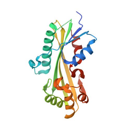 | |
| Ligands 2 Unique | |||||
|---|---|---|---|---|---|
| ID | Chains | Name / Formula / InChI Key | 2D Diagram | 3D Interactions | |
| SO4 Download:Ideal Coordinates CCD File | B [auth A], C [auth A] | SULFATE ION O4 S QAOWNCQODCNURD-UHFFFAOYSA-L |  | ||
| MN Download:Ideal Coordinates CCD File | D [auth A], E [auth A], F [auth A] | MANGANESE (II) ION Mn WAEMQWOKJMHJLA-UHFFFAOYSA-N |  | ||
| Length ( Å ) | Angle ( ˚ ) |
|---|---|
| a = 97.068 | α = 90 |
| b = 97.068 | β = 90 |
| c = 48.894 | γ = 90 |
| Software Name | Purpose |
|---|---|
| SCALA | data scaling |
| SOLVE | phasing |
| RESOLVE | phasing |
| REFMAC | refinement |
| PDB_EXTRACT | data extraction |
| Blu-Ice | data collection |
| CCP4 | data scaling |


