2ZYM | pdb_00002zym

Crystal structure of cyclo/maltodextrin-binding protein complexed with alpha-cyclodextrin


Experimental Data Snapshot

  • Method: X-RAY DIFFRACTION
  • Resolution: 1.80 Å
  • R-Value Free: 
    0.257 (Depositor), 0.276 (DCC) 
  • R-Value Work: 
    0.216 (Depositor), 0.235 (DCC) 
  • R-Value Observed: 
    0.216 (Depositor) 

Starting Model: experimental
View more details

wwPDB Validation 3D Report Full Report

Validation slider image for 2ZYM

This is version 2.1 of the entry. See complete history

Literature

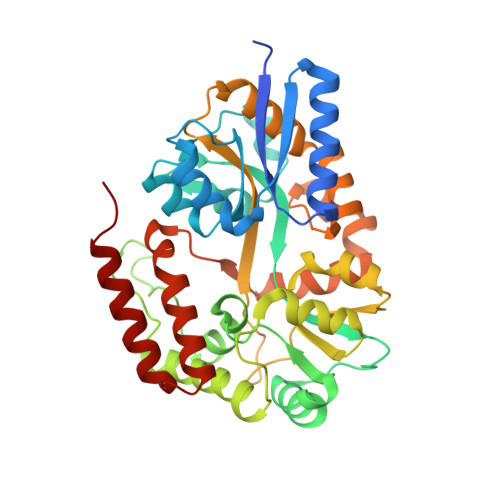
Crystal structures of open and closed forms of cyclo/maltodextrin-binding protein

Matsumoto, N.Yamada, M.Kurakata, Y.Yoshida, H.Kamitori, S.Nishikawa, A.Tonozuka, T.

(2009) FEBS J 276: 3008-3019

  • DOI: https://doi.org/10.1111/j.1742-4658.2009.07020.x
  • Primary Citation Related Structures: 
    2ZYM, 2ZYN, 2ZYO

  • PubMed Abstract: 

    The crystal structures of Thermoactinomyces vulgaris cyclo/maltodextrin-binding protein (TvuCMBP) complexed with alpha-cyclodextrin (alpha-CD), beta-cyclodextrin (beta-CD) and maltotetraose (G4) have been determined. A common functional conformational change among all solute-binding proteins involves switching from an open form to a closed form, which facilitates transporter binding. Escherichia coli maltodextrin-binding protein (EcoMBP), which is structurally homologous to TvuCMBP, has been determined to adopt the open form when complexed with beta-CD and the closed form when bound to G4. Here, we show that, unlike EcoMBP, TvuCMBP-alpha-CD and TvuCMBP-beta-CD adopt the closed form when complexed, whereas TvuCMBP-G4 adopts the open form. Only two glucose residues are evident in the TvuCMBP-G4 structure, and these bind to the C-domain of TvuCMBP in a manner similar to the way in which maltose binds to the C-domain of EcoMBP. The superposition of TvuCMBP-alpha-CD, TvuCMBP-beta-CD and TvuCMBP-gamma-CD shows that the positions and the orientations of three glucose residues in the cyclodextrin molecules overlay remarkably well. In addition, most of the amino acid residues interacting with these three glucose residues also participate in interactions with the two glucose residues in TvuCMBP-G4, regardless of whether the protein is in the closed or open form. Our results suggest that the mechanisms by which TvuCMBP changes from the open to the closed conformation and maintains the closed form appear to be different from those of EcoMBP, despite the fact that the amino acid residues responsible for the initial binding of the ligands are well conserved between TvuCMBP and EcoMBP.


  • Organizational Affiliation
    • Department of Applied Biological Science, Tokyo University of Agriculture and Technology, Japan.

Macromolecule Content 

  • Total Structure Weight: 44.68 kDa 
  • Atom Count: 3,452 
  • Modeled Residue Count: 381 
  • Deposited Residue Count: 397 
  • Unique protein chains: 1

Macromolecules

Find similar proteins by:|  3D Structure
Entity ID: 1
MoleculeChains  Sequence LengthOrganismDetailsImage
Solute-binding protein397Thermoactinomyces vulgarisMutation(s): 0 
UniProt
Find proteins for Q9AJF5 (Thermoactinomyces vulgaris)
Explore Q9AJF5 
Go to UniProtKB:  Q9AJF5
Entity Groups
Sequence Clusters30% Identity50% Identity70% Identity90% Identity95% Identity100% Identity
UniProt GroupQ9AJF5
Sequence Annotations
Expand
Reference Sequence

Oligosaccharides

Help  
Entity ID: 2
MoleculeChains Length2D Diagram GlycosylationD Interactions
Cyclohexakis-(1-4)-(alpha-D-glucopyranose)
B
6N/A
Glycosylation Resources
GlyTouCan: G22307HL
GlyCosmos: G22307HL

Biologically Interesting Molecules (External Reference) 

1 Unique

Experimental Data & Validation

Experimental Data

  • Method: X-RAY DIFFRACTION
  • Resolution: 1.80 Å
  • R-Value Free:  0.257 (Depositor), 0.276 (DCC) 
  • R-Value Work:  0.216 (Depositor), 0.235 (DCC) 
  • R-Value Observed: 0.216 (Depositor) 
Space Group: C 1 2 1
Unit Cell:
Length ( Å )Angle ( ˚ )
a = 83.236α = 90
b = 46.274β = 94.26
c = 85.647γ = 90
Software Package:
Software NamePurpose
CNSrefinement
ADSCdata collection
HKL-2000data reduction
HKL-2000data scaling
MOLREPphasing

Structure Validation

View Full Validation Report


Entry History 

Deposition Data

Revision History  (Full details and data files)

  • Version 1.0: 2009-03-31
    Type: Initial release
  • Version 1.1: 2011-07-13
    Changes: Version format compliance
  • Version 2.0: 2020-07-29
    Type: Remediation
    Reason: Carbohydrate remediation
    Changes: Atomic model, Data collection, Derived calculations, Non-polymer description, Structure summary
  • Version 2.1: 2023-11-01
    Changes: Data collection, Database references, Refinement description, Structure summary