2ZXV | pdb_00002zxv

Crystal structure of putative acetyltransferase from T. thermophilus HB8


Experimental Data Snapshot

  • Method: X-RAY DIFFRACTION
  • Resolution: 2.30 Å
  • R-Value Free: 
    0.266 (Depositor), 0.264 (DCC) 
  • R-Value Work: 
    0.228 (Depositor), 0.228 (DCC) 

Starting Model: experimental
View more details

wwPDB Validation 3D Report Full Report

Validation slider image for 2ZXV

This is version 1.3 of the entry. See complete history

Literature

Genetic Encoding of 3-Iodo-l-Tyrosine in Escherichia coli for Single-Wavelength Anomalous Dispersion Phasing in Protein Crystallography

Sakamoto, K.Murayama, K.Oki, K.Iraha, F.Kato-Murayama, M.Takahashi, M.Ohtake, K.Kobayashi, T.Kuramitsu, S.Shirouzu, M.Yokoyama, S.

(2009) Structure 17: 335-344

  • DOI: https://doi.org/10.1016/j.str.2009.01.008
  • Primary Citation Related Structures: 
    2Z0Z, 2Z10, 2ZXV

  • PubMed Abstract: 

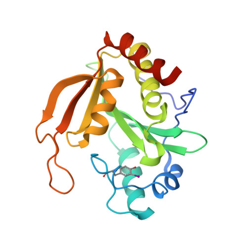
    We developed an Escherichia coli cell-based system to generate proteins containing 3-iodo-L-tyrosine at desired sites, and we used this system for structure determination by single-wavelength anomalous dispersion (SAD) phasing with the strong iodine signal. Tyrosyl-tRNA synthetase from Methanocaldococcus jannaschii was engineered to specifically recognize 3-iodo-L-tyrosine. The 1.7 A crystal structure of the engineered variant, iodoTyrRS-mj, bound with 3-iodo-L-tyrosine revealed the structural basis underlying the strict specificity for this nonnatural substrate; the iodine moiety makes van der Waals contacts with 5 residues at the binding pocket. E. coli cells expressing iodoTyrRS-mj and the suppressor tRNA were used to incorporate 3-iodo-L-tyrosine site specifically into the ribosomal protein N-acetyltransferase from Thermus thermophilus. The crystal structure of this enzyme with iodotyrosine was determined at 1.8 and 2.2 Angstroms resolutions by SAD phasing at CuK alpha and CrK alpha wavelengths, respectively. The native structure, determined by molecular replacement, revealed no significant structural distortion caused by iodotyrosine incorporation.


  • Organizational Affiliation
    • RIKEN Systems and Structural Biology Center, 1-7-22 Suehiro-cho, Tsurumi, Yokohama 230-0045, Japan.

Macromolecule Content 

  • Total Structure Weight: 89.15 kDa 
  • Atom Count: 6,316 
  • Modeled Residue Count: 752 
  • Deposited Residue Count: 776 
  • Unique protein chains: 1

Macromolecules

Find similar proteins by:|  3D Structure
Entity ID: 1
MoleculeChains  Sequence LengthOrganismDetailsImage
Putative uncharacterized protein TTHA1799
A, B, C, D
194Thermus thermophilus HB8Mutation(s): 0 
Gene Names: TTHA1799
EC: 2.3.1.128
UniProt
Find proteins for Q5SHD1 (Thermus thermophilus (strain ATCC 27634 / DSM 579 / HB8))
Explore Q5SHD1 
Go to UniProtKB:  Q5SHD1
Entity Groups
Sequence Clusters30% Identity50% Identity70% Identity90% Identity95% Identity100% Identity
UniProt GroupQ5SHD1
Sequence Annotations
Expand
Reference Sequence

Small Molecules

Modified Residues  1 Unique
IDChains TypeFormula2D DiagramParent
IYR
Query on IYR
A, B, C, D
L-PEPTIDE LINKINGC9 H10 I N O3TYR

Experimental Data & Validation

Experimental Data

  • Method: X-RAY DIFFRACTION
  • Resolution: 2.30 Å
  • R-Value Free:  0.266 (Depositor), 0.264 (DCC) 
  • R-Value Work:  0.228 (Depositor), 0.228 (DCC) 
Space Group: I 4
Unit Cell:
Length ( Å )Angle ( ˚ )
a = 127.8α = 90
b = 127.8β = 90
c = 122.1γ = 90
Software Package:
Software NamePurpose
CNSrefinement
CrystalCleardata collection
HKL-2000data reduction
HKL-2000data scaling
MOLREPphasing

Structure Validation

View Full Validation Report


Entry History 

Revision History  (Full details and data files)

  • Version 1.0: 2009-02-17
    Type: Initial release
  • Version 1.1: 2011-07-13
    Changes: Version format compliance
  • Version 1.2: 2023-11-01
    Changes: Data collection, Database references, Derived calculations, Refinement description
  • Version 1.3: 2024-11-20
    Changes: Structure summary