2ZTW | pdb_00002ztw

Structure of 3-isopropylmalate dehydrogenase in complex with the inhibitor and NAD+


Experimental Data Snapshot

  • Method: X-RAY DIFFRACTION
  • Resolution: 2.79 Å
  • R-Value Free: 
    0.250 (Depositor), 0.247 (DCC) 
  • R-Value Work: 
    0.197 (Depositor), 0.200 (DCC) 
  • R-Value Observed: 
    0.200 (Depositor) 

Starting Model: experimental
View more details

wwPDB Validation 3D Report Full Report

Validation slider image for 2ZTW

Ligand Structure Quality Assessment 


This is version 1.2 of the entry. See complete history

Literature

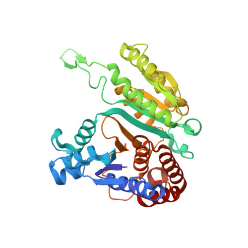
Crystal structure of 3-isopropylmalate dehydrogenase in complex with NAD(+) and a designed inhibitor

Nango, E.Yamamoto, T.Kumasaka, T.Eguchi, T.

(2009) Bioorg Med Chem 17: 7789-7794

  • DOI: https://doi.org/10.1016/j.bmc.2009.09.025
  • Primary Citation Related Structures: 
    2ZTW

  • PubMed Abstract: 

    Isopropylmalate dehydrogenase (IPMDH) is the third enzyme specific to leucine biosynthesis in microorganisms and plants, and catalyzes the oxidative decarboxylation of (2R,3S)-3-isopropylmalate to alpha-ketoisocaproate using NAD(+) as an oxidizing agent. In this study, a thia-analogue of the substrate was designed and synthesized as an inhibitor for IPMDH. The analogue showed strong competitive inhibitory activity with K(i)=62nM toward IPMDH derived from Thermus thermophilus. Moreover, the crystal structure of T. thermophilus IPMDH in a ternary complex with NAD(+) and the inhibitor has been determined at 2.8A resolution. The inhibitor exists as a decarboxylated product with an enol/enolate form in the active site. The product interacts with Arg 94, Asn 102, Ser 259, Glu 270, and a water molecule hydrogen-bonding with Arg 132. All interactions between the product and the enzyme were observed in the position associated with keto-enol tautomerization. This result implies that the tautomerization step of the thia-analogue during the IPMDH reaction is involved in the inhibition.


  • Organizational Affiliation
    • Department of Chemistry, Tokyo Institute of Technology, O-okayama, Meguro-ku, Tokyo 152-8551, Japan.

Macromolecule Content 

  • Total Structure Weight: 37.65 kDa 
  • Atom Count: 2,702 
  • Modeled Residue Count: 345 
  • Deposited Residue Count: 345 
  • Unique protein chains: 1

Macromolecules

Find similar proteins by:|  3D Structure
Entity ID: 1
MoleculeChains  Sequence LengthOrganismDetailsImage
3-isopropylmalate dehydrogenase345Thermus thermophilusMutation(s): 0 
Gene Names: leuBTTHA1230
EC: 1.1.1.85
UniProt
Find proteins for Q5SIY4 (Thermus thermophilus (strain ATCC 27634 / DSM 579 / HB8))
Explore Q5SIY4 
Go to UniProtKB:  Q5SIY4
Entity Groups
Sequence Clusters30% Identity50% Identity70% Identity90% Identity95% Identity100% Identity
UniProt GroupQ5SIY4
Sequence Annotations
Expand
Reference Sequence

Experimental Data & Validation

Experimental Data

  • Method: X-RAY DIFFRACTION
  • Resolution: 2.79 Å
  • R-Value Free:  0.250 (Depositor), 0.247 (DCC) 
  • R-Value Work:  0.197 (Depositor), 0.200 (DCC) 
  • R-Value Observed: 0.200 (Depositor) 
Space Group: P 61 2 2
Unit Cell:
Length ( Å )Angle ( ˚ )
a = 103.507α = 90
b = 103.507β = 90
c = 186.418γ = 120
Software Package:
Software NamePurpose
REFMACrefinement
HKL-2000data collection
HKL-2000data reduction
HKL-2000data scaling
MOLREPphasing

Structure Validation

View Full Validation Report


Ligand Structure Quality Assessment 


Entry History 

Deposition Data

Revision History  (Full details and data files)

  • Version 1.0: 2009-10-13
    Type: Initial release
  • Version 1.1: 2011-07-13
    Changes: Version format compliance
  • Version 1.2: 2023-11-01
    Changes: Data collection, Database references, Derived calculations, Refinement description