2ZSC | pdb_00002zsc

Tamavidin2, Novel Avidin-like Biotin-Binding Proteins from an Edible Mushroom


Experimental Data Snapshot

  • Method: X-RAY DIFFRACTION
  • Resolution: 1.30 Å
  • R-Value Free: 
    0.238 (Depositor), 0.234 (DCC) 
  • R-Value Work: 
    0.199 (Depositor), 0.197 (DCC) 
  • R-Value Observed: 
    0.201 (Depositor) 

Starting Model: experimental
View more details

wwPDB Validation 3D Report Full Report

Validation slider image for 2ZSC

Ligand Structure Quality Assessment 


This is version 1.2 of the entry. See complete history

Literature

Tamavidins--novel avidin-like biotin-binding proteins from the Tamogitake mushroom

Takakura, Y.Tsunashima, M.Suzuki, J.Usami, S.Kakuta, Y.Okino, N.Ito, M.Yamamoto, T.

(2009) FEBS J 276: 1383-1397

  • DOI: https://doi.org/10.1111/j.1742-4658.2009.06879.x
  • Primary Citation Related Structures: 
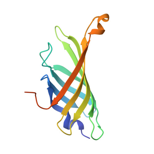
    2ZSC

  • PubMed Abstract: 

    Novel biotin-binding proteins, referred to herein as tamavidin 1 and tamavidin 2, were found in a basidiomycete fungus, Pleurotus cornucopiae, known as the Tamogitake mushroom. These are the first avidin-like proteins to be discovered in organisms other than birds and bacteria. Tamavidin 1 and tamavidin 2 have amino acid sequences with 31% and 36% identity, respectively, to avidin, and 47% and 48% identity, respectively, to streptavidin. Unlike any other biotin-binding proteins, tamavidin 1 and tamavidin 2 are expressed as soluble proteins at a high level in Escherichia coli. Recombinant tamavidin 2 was purified as a tetrameric protein in a single step by 2-iminobiotin affinity chromatography, with a yield of 5 mg per 100 mL culture of E. coli. The kinetic parameters measured by a BIAcore biosensor indicated that recombinant tamavidin 2 binds biotin with high affinity, in a similar manner to binding by avidin and streptavidin. The overall crystal structure of recombinant tamavidin 2 is similar to that of avidin and streptavidin. However, recombinant tamavidin 2 is immunologically distinct from avidin and streptavidin. Tamavidin 2 and streptavidin are very similar in terms of the arrangement of the residues interacting with biotin, but different with regard to the number of hydrogen bonds to biotin carboxylate. Recombinant tamavidin 2 is more stable than avidin and streptavidin at high temperature, and nonspecific binding to DNA and human serum by recombinant tamavidin 2 is lower than that for avidin. These findings highlight tamavidin 2 as a probable powerful tool, in addition to avidin and streptavidin, in numerous applications of biotin-binding proteins.


  • Organizational Affiliation
    • Plant Innovation Center, Japan Tobacco, Inc., Iwata, Shizuoka, Japan. yoshimitsu.takakura@ims.jti.co.jp

Macromolecule Content 

  • Total Structure Weight: 32.35 kDa 
  • Atom Count: 2,600 
  • Modeled Residue Count: 246 
  • Deposited Residue Count: 282 
  • Unique protein chains: 1

Macromolecules

Find similar proteins by:|  3D Structure
Entity ID: 1
MoleculeChains  Sequence LengthOrganismDetailsImage
Tamavidin2
A, B
141Pleurotus cornucopiaeMutation(s): 0 
UniProt
Find proteins for B9A0T7 (Pleurotus cornucopiae)
Explore B9A0T7 
Go to UniProtKB:  B9A0T7
Entity Groups
Sequence Clusters30% Identity50% Identity70% Identity90% Identity95% Identity100% Identity
UniProt GroupB9A0T7
Sequence Annotations
Expand
Reference Sequence

Small Molecules

Ligands 3 Unique
IDChains Name / Formula / InChI Key2D Diagram3D Interactions
BTN

Query on BTN


Download:Ideal Coordinates CCD File
G [auth A],
O [auth B]
BIOTIN
C10 H16 N2 O3 S
YBJHBAHKTGYVGT-ZKWXMUAHSA-N
GOL

Query on GOL


Download:Ideal Coordinates CCD File
C [auth A]
D [auth A]
E [auth A]
F [auth A]
I [auth B]
C [auth A],
D [auth A],
E [auth A],
F [auth A],
I [auth B],
J [auth B],
K [auth B],
L [auth B],
M [auth B]
GLYCEROL
C3 H8 O3
PEDCQBHIVMGVHV-UHFFFAOYSA-N
MG

Query on MG


Download:Ideal Coordinates CCD File
H [auth A],
N [auth B],
P [auth B]
MAGNESIUM ION
Mg
JLVVSXFLKOJNIY-UHFFFAOYSA-N

Experimental Data & Validation

Experimental Data

  • Method: X-RAY DIFFRACTION
  • Resolution: 1.30 Å
  • R-Value Free:  0.238 (Depositor), 0.234 (DCC) 
  • R-Value Work:  0.199 (Depositor), 0.197 (DCC) 
  • R-Value Observed: 0.201 (Depositor) 
Space Group: C 1 2 1
Unit Cell:
Length ( Å )Angle ( ˚ )
a = 78.382α = 90
b = 80.017β = 132.32
c = 55.733γ = 90
Software Package:
Software NamePurpose
REFMACrefinement
HKL-2000data reduction
HKL-2000data scaling
MOLREPphasing

Structure Validation

View Full Validation Report


Ligand Structure Quality Assessment 


Entry History 

Deposition Data

Revision History  (Full details and data files)

  • Version 1.0: 2009-02-17
    Type: Initial release
  • Version 1.1: 2011-07-13
    Changes: Non-polymer description, Version format compliance
  • Version 1.2: 2023-11-01
    Changes: Data collection, Database references, Derived calculations, Refinement description