2ZQ8 | pdb_00002zq8

Apo structure of class a beta-lactamase Toho-1 R274N/R276N double mutant


Experimental Data Snapshot

  • Method: X-RAY DIFFRACTION
  • Resolution: 1.03 Å
  • R-Value Free: 
    0.148 (Depositor), 0.148 (DCC) 
  • R-Value Work: 
    0.138 (Depositor), 0.138 (DCC) 
  • R-Value Observed: 
    0.138 (Depositor) 

Starting Model: experimental
View more details

wwPDB Validation 3D Report Full Report

Validation slider image for 2ZQ8

This is version 1.3 of the entry. See complete history

Literature

Improvement of crystal quality by surface mutations of beta-lactamase Toho-1

Shimamura, T.Nitanai, Y.Uchiyama, T.Matsuzawa, H.

(2009) Acta Crystallogr Sect F Struct Biol Cryst Commun 65: 379-382

  • DOI: https://doi.org/10.1107/S1744309109008240
  • Primary Citation Related Structures: 
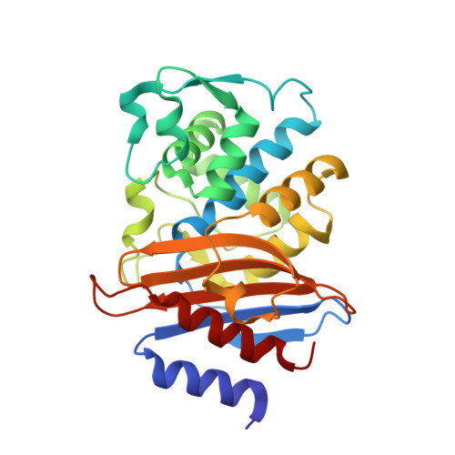
    2ZQ8

  • PubMed Abstract: 

    The beta-lactamase Toho-1 exhibits a strong tendency to form merohedrally twinned crystals. Here, the crystal quality of Toho-1 was improved by using surface modification to remove a sulfate ion involved in crystal packing. The surface-modified Toho-1 variant (R274N/R276N) was crystallized under similar conditions to those used for wild-type Toho-1. R274N/R276N did not form merohedrally twinned crystals. The crystals diffracted to a significantly higher resolution (approximately 0.97 A) than the wild-type crystals (1.65 A); they belonged to the same space group and had almost identical unit-cell parameters to those of wild-type Toho-1.


  • Organizational Affiliation
    • RIKEN SPring-8 Center, Harima Institute, Kouto, Sayo, Hyogo 679-5148, Japan. t.shimamura@mfour.med.kyoto-u.ac.jp

Macromolecule Content 

  • Total Structure Weight: 28.66 kDa 
  • Atom Count: 2,733 
  • Modeled Residue Count: 261 
  • Deposited Residue Count: 262 
  • Unique protein chains: 1

Macromolecules

Find similar proteins by:|  3D Structure
Entity ID: 1
MoleculeChains  Sequence LengthOrganismDetailsImage
Beta-lactamase Toho-1262Escherichia coliMutation(s): 2 
Gene Names: bla
EC: 3.5.2.6
UniProt
Find proteins for Q47066 (Escherichia coli)
Explore Q47066 
Go to UniProtKB:  Q47066
Entity Groups
Sequence Clusters30% Identity50% Identity70% Identity90% Identity95% Identity100% Identity
UniProt GroupQ47066
Sequence Annotations
Expand
Reference Sequence

Experimental Data & Validation

Experimental Data

  • Method: X-RAY DIFFRACTION
  • Resolution: 1.03 Å
  • R-Value Free:  0.148 (Depositor), 0.148 (DCC) 
  • R-Value Work:  0.138 (Depositor), 0.138 (DCC) 
  • R-Value Observed: 0.138 (Depositor) 
Space Group: P 32 2 1
Unit Cell:
Length ( Å )Angle ( ˚ )
a = 72.511α = 90
b = 72.511β = 90
c = 97.477γ = 120
Software Package:
Software NamePurpose
MOLREPphasing
REFMACrefinement
HKL-2000data reduction
SCALEPACKdata scaling

Structure Validation

View Full Validation Report


Entry History 

Deposition Data

Revision History  (Full details and data files)

  • Version 1.0: 2009-07-28
    Type: Initial release
  • Version 1.1: 2011-07-13
    Changes: Version format compliance
  • Version 1.2: 2021-11-10
    Changes: Database references, Derived calculations
  • Version 1.3: 2023-11-01
    Changes: Data collection, Refinement description