2ZPY | pdb_00002zpy

Crystal structure of the mouse radxin FERM domain complexed with the mouse CD44 cytoplasmic peptide


Experimental Data Snapshot

  • Method: X-RAY DIFFRACTION
  • Resolution: 2.10 Å
  • R-Value Free: 
    0.256 (Depositor), 0.255 (DCC) 
  • R-Value Work: 
    0.231 (Depositor), 0.231 (DCC) 

Starting Model: experimental
View more details

wwPDB Validation 3D Report Full Report

Validation slider image for 2ZPY

This is version 1.2 of the entry. See complete history

Literature

Structural basis for CD44 recognition by ERM proteins

Mori, T.Kitano, K.Terawaki, S.Maesaki, R.Fukami, Y.Hakoshima, T.

(2008) J Biological Chem 283: 29602-29612

  • DOI: https://doi.org/10.1074/jbc.M803606200
  • Primary Citation Related Structures: 
    2ZPY

  • PubMed Abstract: 

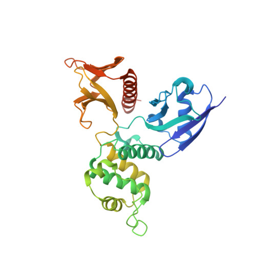
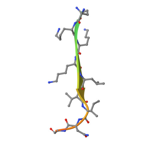
    CD44 is an important adhesion molecule that functions as the major hyaluronan receptor which mediates cell adhesion and migration in a variety of physiological and pathological processes. Although full activity of CD44 requires binding to ERM (ezrin/radixin/moesin) proteins, the CD44 cytoplasmic region, consisting of 72 amino acid residues, lacks the Motif-1 consensus sequence for ERM binding found in intercellular adhesion molecule (ICAM)-2 and other adhesion molecules of the immunoglobulin superfamily. Ultracentrifugation sedimentation studies and circular dichroism measurements revealed an extended monomeric form of the cytoplasmic peptide in solution. The crystal structure of the radixin FERM domain complexed with a CD44 cytoplasmic peptide reveals that the KKKLVIN sequence of the peptide forms a beta strand followed by a short loop structure that binds subdomain C of the FERM domain. Like Motif-1 binding, the CD44 beta strand binds the shallow groove between strand beta5C and helix alpha1C and augments the beta sheet beta5C-beta7C from subdomain C. Two hydrophobic CD44 residues, Leu and Ile, are docked into a hydrophobic pocket with the formation of hydrogen bonds between Asn of the CD44 short loop and loop beta4C-beta5C from subdomain C. This binding mode resembles that of NEP (neutral endopeptidase 24.11) rather than ICAM-2. Our results reveal a characteristic versatility of peptide recognition by the FERM domains from ERM proteins, suggest a possible mechanism by which the CD44 tail is released from the cytoskeleton for nuclear translocation by regulated intramembrane proteolysis, and provide a structural basis for Smad1 interactions with activated CD44 bound to ERM protein.


  • Organizational Affiliation
    • Structural Biology Laboratory, Nara Institute of Science and Technology, 8916-5 Takayama, Ikoma, Nara 630-0192, Japan.

Macromolecule Content 

  • Total Structure Weight: 39.11 kDa 
  • Atom Count: 2,714 
  • Modeled Residue Count: 304 
  • Deposited Residue Count: 332 
  • Unique protein chains: 2

Macromolecules

Find similar proteins by:|  3D Structure
Entity ID: 1
MoleculeChains  Sequence LengthOrganismDetailsImage
Radixin312Mus musculusMutation(s): 0 
Gene Names: Rdx
UniProt & NIH Common Fund Data Resources
Find proteins for P26043 (Mus musculus)
Explore P26043 
Go to UniProtKB:  P26043
IMPC:  MGI:97887
Entity Groups
Sequence Clusters30% Identity50% Identity70% Identity90% Identity95% Identity100% Identity
UniProt GroupP26043
Sequence Annotations
Expand
Reference Sequence
Find similar proteins by:  Sequence   |   3D Structure  
Entity ID: 2
MoleculeChains  Sequence LengthOrganismDetailsImage
CD44 antigen20N/AMutation(s): 0 
UniProt
Find proteins for P15379 (Mus musculus)
Explore P15379 
Go to UniProtKB:  P15379
Entity Groups
Sequence Clusters30% Identity50% Identity70% Identity90% Identity95% Identity100% Identity
UniProt GroupP15379
Sequence Annotations
Expand
Reference Sequence

Experimental Data & Validation

Experimental Data

  • Method: X-RAY DIFFRACTION
  • Resolution: 2.10 Å
  • R-Value Free:  0.256 (Depositor), 0.255 (DCC) 
  • R-Value Work:  0.231 (Depositor), 0.231 (DCC) 
Space Group: P 21 21 21
Unit Cell:
Length ( Å )Angle ( ˚ )
a = 62.7α = 90
b = 66.182β = 90
c = 86.217γ = 90
Software Package:
Software NamePurpose
HKL-2000data collection
PHASERphasing
CNSrefinement
SCALEPACKdata scaling

Structure Validation

View Full Validation Report


Entry History 

Deposition Data

Revision History  (Full details and data files)

  • Version 1.0: 2008-08-26
    Type: Initial release
  • Version 1.1: 2011-07-13
    Changes: Version format compliance
  • Version 1.2: 2023-11-01
    Changes: Data collection, Database references, Refinement description