2ZON | pdb_00002zon

Crystal structure of electron transfer complex of nitrite reductase with cytochrome c


Experimental Data Snapshot

  • Method: X-RAY DIFFRACTION
  • Resolution: 1.70 Å
  • R-Value Free: 
    0.195 (Depositor), 0.194 (DCC) 
  • R-Value Work: 
    0.162 (Depositor), 0.162 (DCC) 
  • R-Value Observed: 
    0.165 (Depositor) 

Starting Models: experimental
View more details

wwPDB Validation 3D Report Full Report

Validation slider image for 2ZON

Ligand Structure Quality Assessment 


This is version 1.3 of the entry. See complete history

Literature

Structural basis of inter-protein electron transfer for nitrite reduction in denitrification

Nojiri, M.Koteishi, H.Nakagami, T.Kobayashi, K.Inoue, T.Yamaguchi, K.Suzuki, S.

(2009) Nature 462: 117-120

  • DOI: https://doi.org/10.1038/nature08507
  • Primary Citation Related Structures: 
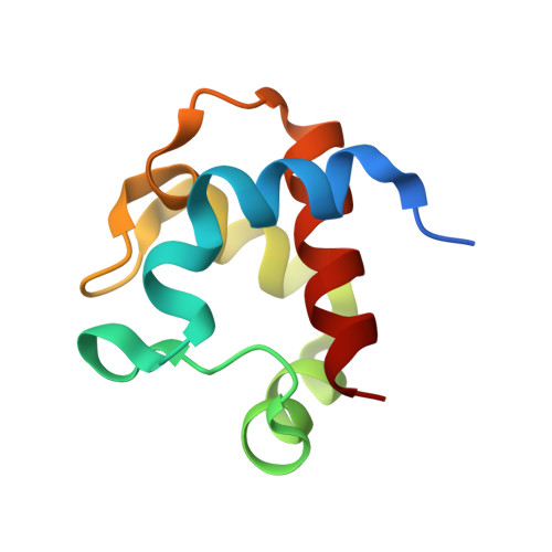
    2ZON

  • PubMed Abstract: 

    Recent earth science studies have pointed out that massive acceleration of the global nitrogen cycle by anthropogenic addition of bio-available nitrogen has led to a host of environmental problems. Nitrous oxide (N(2)O) is a greenhouse gas that is an intermediate during the biological process known as denitrification. Copper-containing nitrite reductase (CuNIR) is a key enzyme in the process; it produces a precursor for N(2)O by catalysing the one-electron reduction of nitrite (NO2-) to nitric oxide (NO). The reduction step is performed by an efficient electron-transfer reaction with a redox-partner protein. However, details of the mechanism during the electron-transfer reaction are still unknown. Here we show the high-resolution crystal structure of the electron-transfer complex for CuNIR with its cognate cytochrome c as the electron donor. The hydrophobic electron-transfer path is formed at the docking interface by desolvation owing to close contact between the two proteins. Structural analysis of the interface highlights an essential role for the loop region with a hydrophobic patch for protein-protein recognition; it also shows how interface construction allows the variation in atomic components to achieve diverse biological electron transfers.


  • Organizational Affiliation
    • Department of Chemistry, Graduate School of Science, Osaka University, Toyonaka, Osaka 560-0043, Japan. nojiri@ch.wani.osaka-u.ac.jp

Macromolecule Content 

  • Total Structure Weight: 119.47 kDa 
  • Atom Count: 9,364 
  • Modeled Residue Count: 1,083 
  • Deposited Residue Count: 1,095 
  • Unique protein chains: 2

Macromolecules

Find similar proteins by:|  3D Structure
Entity ID: 1
MoleculeChains  Sequence LengthOrganismDetailsImage
Dissimilatory copper-containing nitrite reductase
A, B, C
336Achromobacter xylosoxidansMutation(s): 0 
EC: 1.7.2.1
UniProt
Find proteins for O68601 (Alcaligenes xylosoxydans xylosoxydans)
Explore O68601 
Go to UniProtKB:  O68601
Entity Groups
Sequence Clusters30% Identity50% Identity70% Identity90% Identity95% Identity100% Identity
UniProt GroupO68601
Sequence Annotations
Expand
Reference Sequence
Find similar proteins by:|  3D Structure
Entity ID: 2
MoleculeChains  Sequence LengthOrganismDetailsImage
cytochrome c551D [auth G]87Achromobacter xylosoxidansMutation(s): 0 
UniProt
Find proteins for D0VWQ8 (Alcaligenes xylosoxydans xylosoxydans)
Explore D0VWQ8 
Go to UniProtKB:  D0VWQ8
Entity Groups
Sequence Clusters30% Identity50% Identity70% Identity90% Identity95% Identity100% Identity
UniProt GroupD0VWQ8
Sequence Annotations
Expand
Reference Sequence

Experimental Data & Validation

Experimental Data

  • Method: X-RAY DIFFRACTION
  • Resolution: 1.70 Å
  • R-Value Free:  0.195 (Depositor), 0.194 (DCC) 
  • R-Value Work:  0.162 (Depositor), 0.162 (DCC) 
  • R-Value Observed: 0.165 (Depositor) 
Space Group: P 21 21 21
Unit Cell:
Length ( Å )Angle ( ˚ )
a = 63.345α = 90
b = 103.942β = 90
c = 163.144γ = 90
Software Package:
Software NamePurpose
REFMACrefinement
HKL-2000data collection
HKL-2000data reduction
SCALEPACKdata scaling

Structure Validation

View Full Validation Report


Ligand Structure Quality Assessment 


Entry History 

Deposition Data

Revision History  (Full details and data files)

  • Version 1.0: 2009-06-09
    Type: Initial release
  • Version 1.1: 2011-07-13
    Changes: Version format compliance
  • Version 1.2: 2013-09-18
    Changes: Derived calculations
  • Version 1.3: 2023-11-01
    Changes: Data collection, Database references, Derived calculations, Refinement description