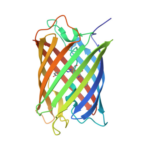
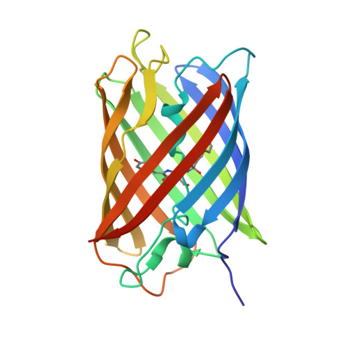
2ZMW

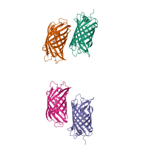
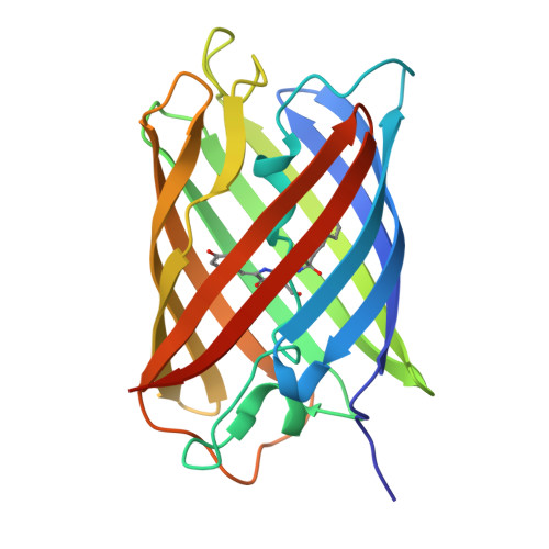
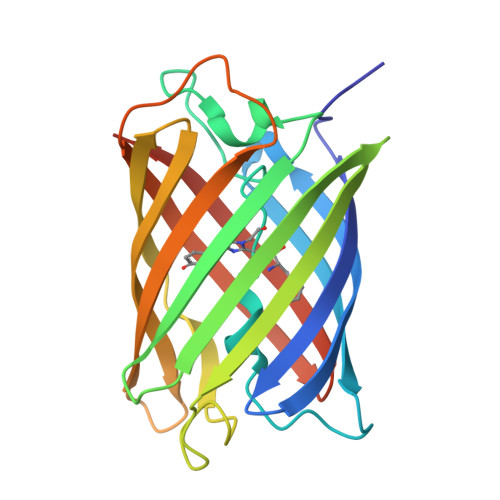
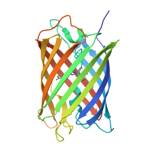
Crystal Structure of Monomeric Kusabira-Orange (MKO), Orange-Emitting GFP-like Protein, at pH 6.0


Experimental Data Snapshot

  • Method: X-RAY DIFFRACTION
  • Resolution: 2.00 Å
  • R-Value Free: 0.232 
  • R-Value Work: 0.197 
  • R-Value Observed: 0.197 

Starting Model: experimental
View more details

wwPDB Validation   3D Report Full Report


This is version 2.1 of the entry. See complete history


Literature

Structural Characterization of a Thiazoline-Containing Chromophore in an Orange Fluorescent Protein, Monomeric Kusabira Orange

Kikuchi, A.Fukumura, E.Karasawa, S.Mizuno, H.Miyawaki, A.Shiro, Y.

(2008) Biochemistry 47: 11573-11580

  • DOI: https://doi.org/10.1021/bi800727v
  • Primary Citation of Related Structures:  
    2ZMU, 2ZMW

  • PubMed Abstract: 

    Monomeric Kusabira Orange (mKO) is a green fluorescent protein (GFP)-like protein that emits orange light at a peak of 559 nm. We analyzed its X-ray structure at 1.65 A and found a novel three-ring chromophore that developed autocatalytically from a Cys65-Tyr66-Glu67 tripeptide in which the side chain of Cys65 formed the third 2-hydroxy-3-thiazoline ring. As a result, the chromophore contained the CNCOH group at the 2-position of the imidazolinone moiety such that the conjugated pi-electron system of the chromophore was more extended than that of GFP but less extended than that of the Discosoma sp. red fluorescent protein (DsRed). Since a sulfur atom has potent nucleophilic character, the third 3-thiazoline ring is rapidly and completely cyclized. Furthermore, our structure reveals the presence of a pi-pi stacking interaction between His197 and the chromophore as well as a pi-cation interaction between Arg69 and the chromophore. These structural findings are sufficient to account for the orange emission, pH tolerance, and photostability of mKO.


  • Organizational Affiliation

    Biometal Science Laboratory, RIKEN SPring-8 Center, 1-1-1, Kouto, Sayo, Hyogo 679-5148, Japan. kikuchi@spring8.or.jp


Macromolecules
Find similar proteins by:  (by identity cutoff)  |  3D Structure
Entity ID: 1
MoleculeChains Sequence LengthOrganismDetailsImage
Fluorescent protein
A, B, C, D
223Lithophyllon concinnaMutation(s): 1 
Gene Names: mKO
UniProt
Find proteins for Q6I7B2 (Lithophyllon concinna)
Explore Q6I7B2 
Go to UniProtKB:  Q6I7B2
Entity Groups  
Sequence Clusters30% Identity50% Identity70% Identity90% Identity95% Identity100% Identity
UniProt GroupQ6I7B2
Sequence Annotations
Expand
  • Reference Sequence
Small Molecules
Modified Residues  1 Unique
IDChains TypeFormula2D DiagramParent
CFY
Query on CFY
A, B, C, D
L-PEPTIDE LINKINGC23 H22 N4 O5 SPHE, CYS, TYR, GLY
Experimental Data & Validation

Experimental Data

  • Method: X-RAY DIFFRACTION
  • Resolution: 2.00 Å
  • R-Value Free: 0.232 
  • R-Value Work: 0.197 
  • R-Value Observed: 0.197 
  • Space Group: P 1 21 1
Unit Cell:
Length ( Å )Angle ( ˚ )
a = 92.57α = 90
b = 72.11β = 102.72
c = 90.27γ = 90
Software Package:
Software NamePurpose
MOLREPphasing
CNSrefinement
ADSCdata collection
HKL-2000data reduction
HKL-2000data scaling

Structure Validation

View Full Validation Report


Entry History 

Deposition Data

Revision History  (Full details and data files)

  • Version 1.0: 2008-10-21
    Type: Initial release
  • Version 1.1: 2011-07-13
    Changes: Version format compliance
  • Version 1.2: 2012-05-02
    Changes: Other, Source and taxonomy
  • Version 1.3: 2023-11-01
    Changes: Data collection, Database references, Derived calculations, Refinement description
  • Version 2.0: 2023-11-15
    Changes: Atomic model, Data collection, Derived calculations
  • Version 2.1: 2024-10-16
    Changes: Structure summary