2ZHZ | pdb_00002zhz

Crystal structure of a pduO-type ATP:cobalamin adenosyltransferase from Burkholderia thailandensis


Experimental Data Snapshot

  • Method: X-RAY DIFFRACTION
  • Resolution: 1.80 Å
  • R-Value Free: 
    0.229 (Depositor), 0.229 (DCC) 
  • R-Value Work: 
    0.201 (Depositor), 0.201 (DCC) 
  • R-Value Observed: 
    0.211 (Depositor) 

wwPDB Validation 3D Report Full Report

Validation slider image for 2ZHZ

Ligand Structure Quality Assessment 


This is version 1.3 of the entry. See complete history

Literature

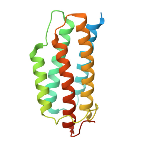
Crystal structure of a PduO-type ATP:cobalamin adenosyltransferase from Burkholderia thailandensis.

Moon, J.H.Park, A.K.Jang, E.H.Kim, H.S.Chi, Y.M.

(2008) Proteins 72: 1066-1070

Macromolecule Content 

  • Total Structure Weight: 59.91 kDa 
  • Atom Count: 4,038 
  • Modeled Residue Count: 494 
  • Deposited Residue Count: 549 
  • Unique protein chains: 1

Macromolecules

Find similar proteins by:|  3D Structure
Entity ID: 1
MoleculeChains  Sequence LengthOrganismDetailsImage
ATP:cob(I)alamin adenosyltransferase, putative
A, B, C
183Burkholderia thailandensisMutation(s): 0 
Gene Names: pduO
EC: 2.5.1.17 (PDB Primary Data), 2.5.1 (UniProt)
UniProt
Find proteins for Q2SZ09 (Burkholderia thailandensis (strain ATCC 700388 / DSM 13276 / CCUG 48851 / CIP 106301 / E264))
Explore Q2SZ09 
Go to UniProtKB:  Q2SZ09
Entity Groups
Sequence Clusters30% Identity50% Identity70% Identity90% Identity95% Identity100% Identity
UniProt GroupQ2SZ09
Sequence Annotations
Expand
Reference Sequence

Small Molecules

Ligands 3 Unique
IDChains Name / Formula / InChI Key2D Diagram3D Interactions
ATP

Query on ATP


Download:Ideal Coordinates CCD File
G [auth B],
H [auth B]
ADENOSINE-5'-TRIPHOSPHATE
C10 H16 N5 O13 P3
ZKHQWZAMYRWXGA-KQYNXXCUSA-N
GOL

Query on GOL


Download:Ideal Coordinates CCD File
E [auth A],
I [auth B],
J [auth C]
GLYCEROL
C3 H8 O3
PEDCQBHIVMGVHV-UHFFFAOYSA-N
MG

Query on MG


Download:Ideal Coordinates CCD File
D [auth A],
F [auth B]
MAGNESIUM ION
Mg
JLVVSXFLKOJNIY-UHFFFAOYSA-N

Experimental Data & Validation

Experimental Data

  • Method: X-RAY DIFFRACTION
  • Resolution: 1.80 Å
  • R-Value Free:  0.229 (Depositor), 0.229 (DCC) 
  • R-Value Work:  0.201 (Depositor), 0.201 (DCC) 
  • R-Value Observed: 0.211 (Depositor) 
Space Group: C 2 2 21
Unit Cell:
Length ( Å )Angle ( ˚ )
a = 53.907α = 90
b = 148.174β = 90
c = 158.096γ = 90
Software Package:
Software NamePurpose
DENZOdata reduction
SCALEPACKdata scaling
CNSrefinement
PDB_EXTRACTdata extraction
HKL-2000data collection
HKL-2000data reduction
CNSphasing

Structure Validation

View Full Validation Report


Ligand Structure Quality Assessment 


Entry History 

Deposition Data

Revision History  (Full details and data files)

  • Version 1.0: 2008-07-29
    Type: Initial release
  • Version 1.1: 2011-07-13
    Changes: Non-polymer description, Version format compliance
  • Version 1.2: 2017-10-11
    Changes: Refinement description
  • Version 1.3: 2024-03-13
    Changes: Data collection, Database references, Derived calculations