2ZHM

Crystal structure of human galectin-9 N-terminal CRD in complex with N-acetyllactosamine trimer (crystal 1)


Experimental Data Snapshot

  • Method: X-RAY DIFFRACTION
  • Resolution: 1.84 Å
  • R-Value Free: 
    0.235 (Depositor), 0.230 (DCC) 
  • R-Value Work: 
    0.182 (Depositor), 0.180 (DCC) 
  • R-Value Observed: 
    0.185 (Depositor) 

wwPDB Validation   3D Report Full Report


Ligand Structure Quality Assessment 


This is version 2.1 of the entry. See complete history


Literature

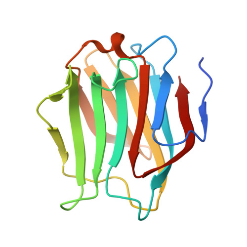
Structural analysis of the recognition mechanism of poly-N-acetyllactosamine by the human galectin-9 N-terminal carbohydrate recognition domain.

Nagae, M.Nishi, N.Murata, T.Usui, T.Nakamura, T.Wakatsuki, S.Kato, R.

(2009) Glycobiology 19: 112-117

  • DOI: https://doi.org/10.1093/glycob/cwn121
  • Primary Citation of Related Structures:  
    2ZHK, 2ZHL, 2ZHM, 2ZHN

  • PubMed Abstract: 

    Galectins are a family of beta-galactoside-specific lectins bearing a conserved carbohydrate recognition domain. Interactions between galectins and poly-N-acetyllactosamine sequences are critical in a variety of biological processes. Galectin-9, a member of the galectin family, has two carbohydrate recognition domains at both the N- and C-terminal regions. Here we report the crystal structure of the human galectin-9 N-terminal carbohydrate recognition domain in complex with N-acetyllactosamine dimers and trimers. These complex structures revealed that the galectin-9 N-terminal carbohydrate recognition domain can recognize internal N-acetyllactosamine units within poly-N-acetyllactosamine chains. Based on these complex structures, we propose two putative recognition modes for poly-N-acetyllactosamine binding by galectins.


  • Organizational Affiliation

    Structural Biology Research Center, Photon Factory, Institute of Materials Structure Science, High Energy Accelerator Research Organization (KEK), Tsukuba, Ibaraki, Japan.


Macromolecules
Find similar proteins by:  (by identity cutoff)  |  3D Structure
Entity ID: 1
MoleculeChains Sequence LengthOrganismDetailsImage
Galectin-9
A, B, C, D
148Homo sapiensMutation(s): 0 
UniProt & NIH Common Fund Data Resources
Find proteins for O00182 (Homo sapiens)
Explore O00182 
Go to UniProtKB:  O00182
PHAROS:  O00182
GTEx:  ENSG00000168961 
Entity Groups  
Sequence Clusters30% Identity50% Identity70% Identity90% Identity95% Identity100% Identity
UniProt GroupO00182
Sequence Annotations
Expand
  • Reference Sequence
Oligosaccharides

Help

Entity ID: 2
MoleculeChains Length2D Diagram Glycosylation3D Interactions
beta-D-galactopyranose-(1-4)-2-acetamido-2-deoxy-beta-D-glucopyranose-(1-3)-beta-D-galactopyranose-(1-4)-2-acetamido-2-deoxy-beta-D-glucopyranose-(1-3)-beta-D-galactopyranose-(1-4)-2-acetamido-2-deoxy-beta-D-glucopyranose
E, F
6N/A
Glycosylation Resources
GlyTouCan:  G46055MA
GlyCosmos:  G46055MA
GlyGen:  G46055MA
Entity ID: 3
MoleculeChains Length2D Diagram Glycosylation3D Interactions
beta-D-galactopyranose-(1-4)-2-acetamido-2-deoxy-beta-D-glucopyranose-(1-3)-beta-D-galactopyranose-(1-4)-2-acetamido-2-deoxy-beta-D-glucopyranose-(1-3)-beta-D-galactopyranose
G
5N/A
Glycosylation Resources
GlyTouCan:  G04165AB
GlyCosmos:  G04165AB
GlyGen:  G04165AB
Entity ID: 4
MoleculeChains Length2D Diagram Glycosylation3D Interactions
beta-D-galactopyranose-(1-4)-2-acetamido-2-deoxy-beta-D-glucopyranose-(1-3)-beta-D-galactopyranose-(1-4)-2-acetamido-2-deoxy-beta-D-glucopyranose
H
4N/A
Glycosylation Resources
GlyTouCan:  G00058MO
GlyCosmos:  G00058MO
GlyGen:  G00058MO
Experimental Data & Validation

Experimental Data

  • Method: X-RAY DIFFRACTION
  • Resolution: 1.84 Å
  • R-Value Free:  0.235 (Depositor), 0.230 (DCC) 
  • R-Value Work:  0.182 (Depositor), 0.180 (DCC) 
  • R-Value Observed: 0.185 (Depositor) 
Space Group: P 21 21 21
Unit Cell:
Length ( Å )Angle ( ˚ )
a = 35.217α = 90
b = 67.964β = 90
c = 219.964γ = 90
Software Package:
Software NamePurpose
MOLREPphasing
REFMACrefinement
HKL-2000data reduction
HKL-2000data scaling

Structure Validation

View Full Validation Report


Ligand Structure Quality Assessment 


Entry History 

Deposition Data

Revision History  (Full details and data files)

  • Version 1.0: 2008-12-30
    Type: Initial release
  • Version 1.1: 2011-07-13
    Changes: Non-polymer description, Version format compliance
  • Version 2.0: 2020-07-29
    Type: Remediation
    Reason: Carbohydrate remediation
    Changes: Advisory, Atomic model, Data collection, Derived calculations, Structure summary
  • Version 2.1: 2024-03-13
    Changes: Data collection, Database references, Structure summary