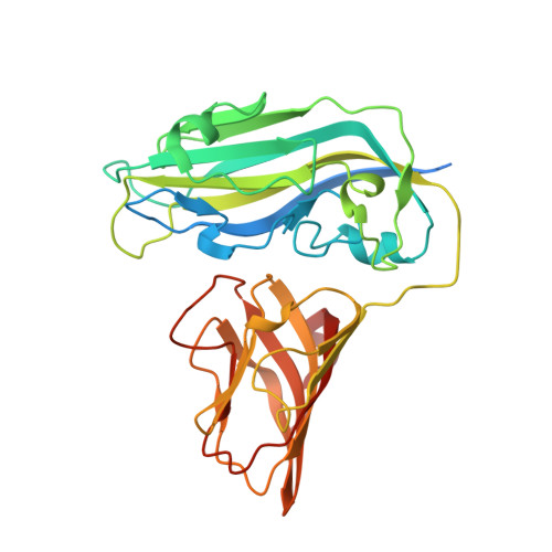
X-ray structure of Melon necrotic spot virus
Wada, Y., Tanaka, H., Yamashita, E., Kubo, C., Ichiki-Uehara, T., Nakazono-Nagaoka, E., Omura, T., Tsukihara, T.(2008) Acta Crystallogr Sect F Struct Biol Cryst Commun 64: 8-13
- PubMed: 18097092 Search on PubMedSearch on PubMed Central
- DOI: https://doi.org/10.1107/S1744309107066481
- Primary Citation Related Structures:
2ZAH - PubMed Abstract:
The structure of melon necrotic spot virus (MNSV) was determined at 2.8 A resolution. Although MNSV is classified into the genus Carmovirus of the family Tombusviridae, the three-dimensional structure of MNSV showed a higher degree of similarity to tomato bushy stunt virus (TBSV), which belongs to the genus Tombusvirus, than to carnation mottle virus (CMtV), turnip crinkle virus (TCV) or cowpea mottle virus (CPMtV) from the genus Carmovirus. Thus, the classification of the family Tombusviridae at the genus level conflicts with the patterns of similarity among coat-protein structures. MNSV is one of the viruses belonging to the genera Tombusvirus or Carmovirus that are naturally transmitted in the soil by zoospores of fungal vectors. The X-ray structure of MNSV provides us with a representative structure of viruses transmitted by fungi.
- Institute for Protein Research, Osaka University, 3-2 Yamada-oka, Suita, Osaka 565-0871, Japan.
Organizational Affiliation: