2Z8X | pdb_00002z8x

Crystal structure of extracellular lipase from Pseudomonas sp. MIS38


Experimental Data Snapshot

  • Method: X-RAY DIFFRACTION
  • Resolution: 1.48 Å
  • R-Value Free: 
    0.194 (Depositor), 0.193 (DCC) 
  • R-Value Work: 
    0.176 (Depositor), 0.174 (DCC) 
  • R-Value Observed: 
    0.177 (Depositor) 

wwPDB Validation 3D Report Full Report

Validation slider image for 2Z8X

This is version 1.2 of the entry. See complete history

Literature

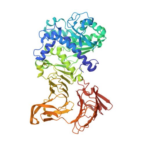
Crystal structure of a family I.3 lipase from Pseudomonas sp. MIS38 in a closed conformation

Angkawidjaja, C.You, D.J.Matsumura, H.Kuwahara, K.Koga, Y.Takano, K.Kanaya, S.

(2007) FEBS Lett 581: 5060-5064

  • DOI: https://doi.org/10.1016/j.febslet.2007.09.048
  • Primary Citation Related Structures: 
    2Z8X, 2Z8Z

  • PubMed Abstract: 

    The crystal structure of a family I.3 lipase from Pseudomonas sp. MIS38 in a closed conformation was determined at 1.5A resolution. This structure highly resembles that of Serratia marcescens LipA in an open conformation, except for the structures of two lids. Lid1 is anchored by a Ca2+ ion (Ca1) in an open conformation, but lacks this Ca1 site and greatly changes its structure and position in a closed conformation. Lid2 forms a helical hairpin in an open conformation, but does not form it and covers the active site in a closed conformation. Based on these results, we discuss on the lid-opening mechanism.


  • Organizational Affiliation
    • Department of Material and Life Science, Graduate School of Engineering, Osaka University, 2-1 Yamadaoka, Suita, Osaka 565-0871, Japan.

Macromolecule Content 

  • Total Structure Weight: 65.1 kDa 
  • Atom Count: 5,334 
  • Modeled Residue Count: 616 
  • Deposited Residue Count: 617 
  • Unique protein chains: 1

Macromolecules

Find similar proteins by:|  3D Structure
Entity ID: 1
MoleculeChains  Sequence LengthOrganismDetailsImage
Lipase617Pseudomonas sp. MIS38Mutation(s): 0 
EC: 3.1.1.3
UniProt
Find proteins for Q9RBY1 (Pseudomonas sp. MIS38)
Explore Q9RBY1 
Go to UniProtKB:  Q9RBY1
Entity Groups
Sequence Clusters30% Identity50% Identity70% Identity90% Identity95% Identity100% Identity
UniProt GroupQ9RBY1
Sequence Annotations
Expand
Reference Sequence

Small Molecules

Ligands 2 Unique
IDChains Name / Formula / InChI Key2D Diagram3D Interactions
ZN

Query on ZN


Download:Ideal Coordinates CCD File
L [auth A],
M [auth A]
ZINC ION
Zn
PTFCDOFLOPIGGS-UHFFFAOYSA-N
CA

Query on CA


Download:Ideal Coordinates CCD File
B [auth A]
C [auth A]
D [auth A]
E [auth A]
F [auth A]
B [auth A],
C [auth A],
D [auth A],
E [auth A],
F [auth A],
G [auth A],
H [auth A],
I [auth A],
J [auth A],
K [auth A]
CALCIUM ION
Ca
BHPQYMZQTOCNFJ-UHFFFAOYSA-N

Experimental Data & Validation

Experimental Data

  • Method: X-RAY DIFFRACTION
  • Resolution: 1.48 Å
  • R-Value Free:  0.194 (Depositor), 0.193 (DCC) 
  • R-Value Work:  0.176 (Depositor), 0.174 (DCC) 
  • R-Value Observed: 0.177 (Depositor) 
Space Group: P 1 21 1
Unit Cell:
Length ( Å )Angle ( ˚ )
a = 49.972α = 90
b = 84.3β = 96.32
c = 86.849γ = 90
Software Package:
Software NamePurpose
REFMACrefinement
HKL-2000data collection
HKL-2000data reduction
HKL-2000data scaling
SHELXSphasing

Structure Validation

View Full Validation Report


Entry History 

Deposition Data

Revision History  (Full details and data files)

  • Version 1.0: 2007-10-30
    Type: Initial release
  • Version 1.1: 2011-07-13
    Changes: Version format compliance
  • Version 1.2: 2024-03-13
    Changes: Data collection, Database references, Derived calculations