2Z6I | pdb_00002z6i

Crystal Structure of S. pneumoniae Enoyl-Acyl Carrier Protein Reductase (FabK)


Experimental Data Snapshot

  • Method: X-RAY DIFFRACTION
  • Resolution: 1.70 Å
  • R-Value Free: 
    0.235 (Depositor), 0.235 (DCC) 
  • R-Value Work: 
    0.202 (Depositor), 0.203 (DCC) 
  • R-Value Observed: 
    0.204 (Depositor) 

wwPDB Validation 3D Report Full Report

Validation slider image for 2Z6I

Ligand Structure Quality Assessment 


This is version 1.2 of the entry. See complete history

Literature

Crystal structure of enoyl-acyl carrier protein reductase (FabK) from Streptococcus pneumoniae reveals the binding mode of an inhibitor.

Saito, J.Yamada, M.Watanabe, T.Iida, M.Kitagawa, H.Takahata, S.Ozawa, T.Takeuchi, Y.Ohsawa, F.

(2008) Protein Sci 17: 691-699

  • DOI: https://doi.org/10.1110/ps.073288808
  • Primary Citation Related Structures: 
    2Z6I, 2Z6J

  • PubMed Abstract: 

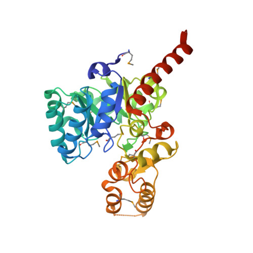
    Enoyl-acyl carrier protein (ACP) reductases are critical for bacterial type II fatty acid biosynthesis and thus are attractive targets for developing novel antibiotics. We determined the crystal structure of enoyl-ACP reductase (FabK) from Streptococcus pneumoniae at 1.7 A resolution. There was one dimer per asymmetric unit. Each subunit formed a triose phosphate isomerase (TIM) barrel structure, and flavin mononucleotide (FMN) was bound as a cofactor in the active site. The overall structure was similar to the enoyl-ACP reductase (ER) of fungal fatty acid synthase and to 2-nitropropane dioxygenase (2-ND) from Pseudomonas aeruginosa, although there were some differences among these structures. We determined the crystal structure of FabK in complex with a phenylimidazole derivative inhibitor to envision the binding site interactions. The crystal structure reveals that the inhibitor binds to a hydrophobic pocket in the active site of FabK, and this is accompanied by induced-fit movements of two loop regions. The thiazole ring and part of the ureido moiety of the inhibitor are involved in a face-to-face pi-pi stacking interaction with the isoalloxazine ring of FMN. The side-chain conformation of the proposed catalytic residue, His144, changes upon complex formation. Lineweaver-Burk plots indicate that the inhibitor binds competitively with respect to NADH, and uncompetitively with respect to crotonoyl coenzyme A. We propose that the primary basis of the inhibitory activity is competition with NADH for binding to FabK, which is the first step of the two-step ping-pong catalytic mechanism.


  • Organizational Affiliation
    • Pharmaceutical Research Center, Meiji Seika Kaisha, Ltd., Kohoku-ku, Yokohama 222-8567, Japan. jun_saito@meiji.co.jp

Macromolecule Content 

  • Total Structure Weight: 72.79 kDa 
  • Atom Count: 5,170 
  • Modeled Residue Count: 638 
  • Deposited Residue Count: 664 
  • Unique protein chains: 1

Macromolecules

Find similar proteins by:|  3D Structure
Entity ID: 1
MoleculeChains  Sequence LengthOrganismDetailsImage
Trans-2-enoyl-ACP reductase II
A, B
332Streptococcus pneumoniaeMutation(s): 0 
Gene Names: fabK
EC: 1.3.1.9
UniProt
Find proteins for Q9FBC5 (Streptococcus pneumoniae)
Explore Q9FBC5 
Go to UniProtKB:  Q9FBC5
Entity Groups
Sequence Clusters30% Identity50% Identity70% Identity90% Identity95% Identity100% Identity
UniProt GroupQ9FBC5
Sequence Annotations
Expand
Reference Sequence

Small Molecules

Ligands 3 Unique
IDChains Name / Formula / InChI Key2D Diagram3D Interactions
FMN

Query on FMN


Download:Ideal Coordinates CCD File
F [auth A],
K [auth B]
FLAVIN MONONUCLEOTIDE
C17 H21 N4 O9 P
FVTCRASFADXXNN-SCRDCRAPSA-N
MPD

Query on MPD


Download:Ideal Coordinates CCD File
G [auth A](4S)-2-METHYL-2,4-PENTANEDIOL
C6 H14 O2
SVTBMSDMJJWYQN-YFKPBYRVSA-N
CA

Query on CA


Download:Ideal Coordinates CCD File
C [auth A]
D [auth A]
E [auth A]
H [auth B]
I [auth B]
C [auth A],
D [auth A],
E [auth A],
H [auth B],
I [auth B],
J [auth B]
CALCIUM ION
Ca
BHPQYMZQTOCNFJ-UHFFFAOYSA-N
Modified Residues  1 Unique
IDChains TypeFormula2D DiagramParent
MSE
Query on MSE
A, B
L-PEPTIDE LINKINGC5 H11 N O2 SeMET

Experimental Data & Validation

Experimental Data

  • Method: X-RAY DIFFRACTION
  • Resolution: 1.70 Å
  • R-Value Free:  0.235 (Depositor), 0.235 (DCC) 
  • R-Value Work:  0.202 (Depositor), 0.203 (DCC) 
  • R-Value Observed: 0.204 (Depositor) 
Space Group: P 1 21 1
Unit Cell:
Length ( Å )Angle ( ˚ )
a = 50.386α = 90
b = 126.895β = 111.54
c = 53.335γ = 90
Software Package:
Software NamePurpose
REFMACrefinement
BSSdata collection
CrystalCleardata reduction
CrystalCleardata scaling
SOLVEphasing

Structure Validation

View Full Validation Report


Ligand Structure Quality Assessment 


Entry History 

Deposition Data

Revision History  (Full details and data files)

  • Version 1.0: 2008-04-01
    Type: Initial release
  • Version 1.1: 2011-07-13
    Changes: Version format compliance
  • Version 1.2: 2024-11-20
    Changes: Data collection, Database references, Derived calculations, Structure summary