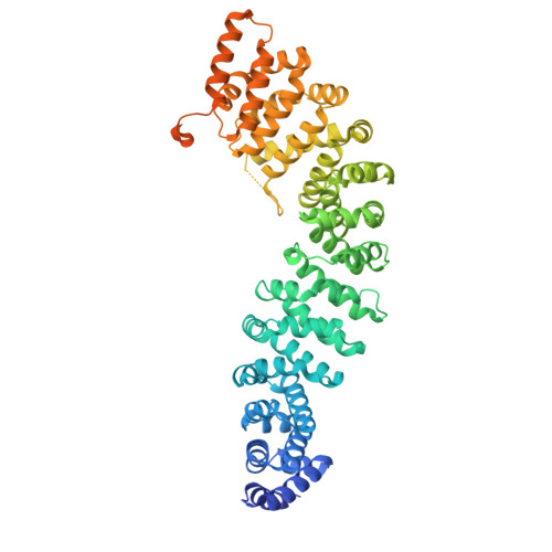
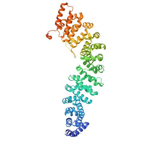
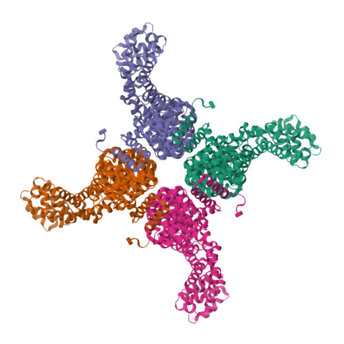
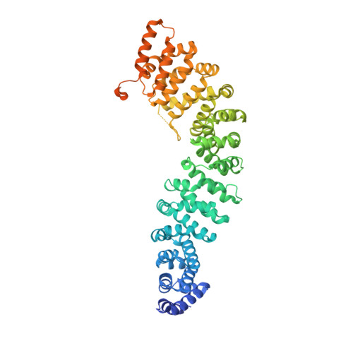
2Z6H

Crystal Structure of Beta-Catenin Armadillo Repeat Region and Its C-Terminal domain


Experimental Data Snapshot

  • Method: X-RAY DIFFRACTION
  • Resolution: 2.20 Å
  • R-Value Free: 0.249 
  • R-Value Work: 0.213 
  • R-Value Observed: 0.213 

wwPDB Validation   3D Report Full Report


This is version 1.2 of the entry. See complete history


Literature

Crystal Structure of a Full-Length beta-Catenin

Xing, Y.Takemaru, K.Liu, J.Berndt, J.D.Zheng, J.J.Moon, R.T.Xu, W.

(2008) Structure 16: 478-487

  • DOI: https://doi.org/10.1016/j.str.2007.12.021
  • Primary Citation of Related Structures:  
    2Z6G, 2Z6H

  • PubMed Abstract: 

    beta-catenin plays essential roles in cell adhesion and Wnt signaling, while deregulation of beta-catenin is associated with multiple diseases including cancers. Here, we report the crystal structures of full-length zebrafish beta-catenin and a human beta-catenin fragment that contains both the armadillo repeat and the C-terminal domains. Our structures reveal that the N-terminal region of the C-terminal domain, a key component of the C-terminal transactivation domain, forms a long alpha helix that packs on the C-terminal end of the armadillo repeat domain, and thus forms part of the beta-catenin superhelical core. The existence of this helix redefines our view of interactions of beta-catenin with some of its critical partners, including ICAT and Chibby, which may form extensive interactions with this C-terminal domain alpha helix. Our crystallographic and NMR studies also suggest that the unstructured N-terminal and C-terminal tails interact with the ordered armadillo repeat domain in a dynamic and variable manner.


  • Organizational Affiliation

    Department of Biological Structure, University of Washington School of Medicine, Seattle, WA 98195, USA.


Macromolecules
Find similar proteins by:  (by identity cutoff)  |  3D Structure
Entity ID: 1
MoleculeChains Sequence LengthOrganismDetailsImage
Catenin beta-1644Homo sapiensMutation(s): 0 
UniProt & NIH Common Fund Data Resources
Find proteins for P35222 (Homo sapiens)
Explore P35222 
Go to UniProtKB:  P35222
PHAROS:  P35222
GTEx:  ENSG00000168036 
Entity Groups  
Sequence Clusters30% Identity50% Identity70% Identity90% Identity95% Identity100% Identity
UniProt GroupP35222
Sequence Annotations
Expand
  • Reference Sequence
Experimental Data & Validation

Experimental Data

  • Method: X-RAY DIFFRACTION
  • Resolution: 2.20 Å
  • R-Value Free: 0.249 
  • R-Value Work: 0.213 
  • R-Value Observed: 0.213 
  • Space Group: I 4 2 2
Unit Cell:
Length ( Å )Angle ( ˚ )
a = 104.229α = 90
b = 104.229β = 90
c = 259.391γ = 90
Software Package:
Software NamePurpose
SOLVEphasing
CNSrefinement
HKL-2000data reduction
SCALEPACKdata scaling

Structure Validation

View Full Validation Report


Entry History 

Deposition Data

Revision History  (Full details and data files)

  • Version 1.0: 2008-02-12
    Type: Initial release
  • Version 1.1: 2011-07-13
    Changes: Version format compliance
  • Version 1.2: 2024-03-13
    Changes: Data collection, Database references