2Z6A | pdb_00002z6a

S-Adenosyl-L-methionine-Dependent Methyl Transfer: Observable Precatalytic Intermediates during DNA Cytosine Methylation


Experimental Data Snapshot

  • Method: X-RAY DIFFRACTION
  • Resolution: 2.88 Å
  • R-Value Free: 
    0.280 (Depositor) 
  • R-Value Work: 
    0.220 (Depositor), 0.229 (DCC) 
  • R-Value Observed: 
    0.239 (Depositor) 

Starting Model: experimental
View more details

wwPDB Validation 3D Report Full Report

Validation slider image for 2Z6A

Ligand Structure Quality Assessment 


This is version 1.4 of the entry. See complete history

Literature

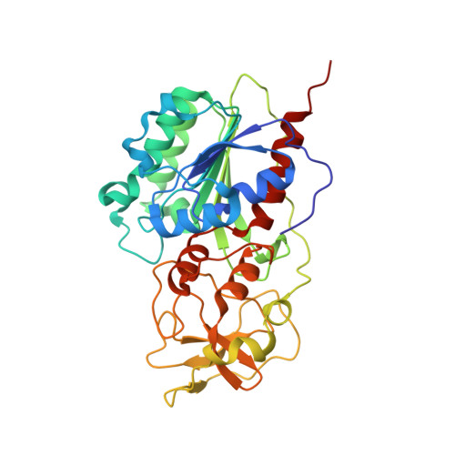
S-Adenosyl-l-methionine-Dependent Methyl Transfer: Observable Precatalytic Intermediates during DNA Cytosine Methylation

Youngblood, B.Shieh, F.K.Buller, F.Bullock, T.Reich, N.O.

(2007) Biochemistry 46: 8766-8775

  • DOI: https://doi.org/10.1021/bi7005948
  • Primary Citation Related Structures: 
    2Z6A

  • PubMed Abstract: 

    S-adenosyl-L-methionine- (AdoMet-) dependent methyltransferases are widespread, play critical roles in diverse biological pathways, and are antibiotic and cancer drug targets. Presently missing from our understanding of any AdoMet-dependent methyl-transfer reaction is a high-resolution structure of a precatalytic enzyme/AdoMet/DNA complex. The catalytic mechanism of DNA cytosine methylation was studied by structurally and functionally characterizing several active site mutants of the bacterial enzyme M.HhaI. The 2.64 A resolution protein/DNA/AdoMet structure of the inactive C81A M.HhaI mutant suggests that active site water, an approximately 13 degree tilt of the target base toward the active site nucleophile, and the presence or absence of the cofactor methylsulfonium are coupled via a hydrogen-bonding network involving Tyr167. The active site in the mutant complex is assembled to optimally align the pyrimidine for nucleophilic attack and subsequent methyl transfer, consistent with previous molecular dynamics ab initio and quantum mechanics/molecular mechanics calculations. The mutant/DNA/AdoHcy structure (2.88 A resolution) provides a direct comparison to the postcatalytic complex. A third C81A ternary structure (2.22 A resolution) reveals hydrolysis of AdoMet to adenosine in the active site, further validating the coupling between the methionine portion of AdoMet and ultimately validating the structural observation of a prechemistry/postchemistry water network. Disruption of this hydrogen-bonding network by a Tyr167 to Phe167 mutation does not alter the kinetics of nucleophilic attack or methyl transfer. However, the Y167F mutant shows detectable changes in kcat, caused by the perturbed kinetics of AdoHcy release. These results provide a basis for including an extensive hydrogen-bonding network in controlling the rate-limiting product release steps during cytosine methylation.


  • Organizational Affiliation
    • Program in Biomolecular Science and Engineering, University of California, Santa Barbara, California 93106-9510, USA.

Macromolecule Content 

  • Total Structure Weight: 45.02 kDa 
  • Atom Count: 3,192 
  • Modeled Residue Count: 352 
  • Deposited Residue Count: 352 
  • Unique protein chains: 1
  • Unique nucleic acid chains: 2

Macromolecules


Find similar proteins by:|  3D Structure
Entity ID: 3
MoleculeChains  Sequence LengthOrganismDetailsImage
Modification methylase HhaIC [auth A]327Haemophilus parahaemolyticusMutation(s): 1 
Gene Names: hhaIM
EC: 2.1.1.37
UniProt
Find proteins for P05102 (Haemophilus parahaemolyticus)
Explore P05102 
Go to UniProtKB:  P05102
Entity Groups
Sequence Clusters30% Identity50% Identity70% Identity90% Identity95% Identity100% Identity
UniProt GroupP05102
Sequence Annotations
Expand
Reference Sequence
Find similar nucleic acids by:  Sequence
Entity ID: 1
MoleculeChains LengthOrganismImage
DNA (5'-D(*DGP*DAP*DTP*DAP*DGP*DCP*DGP*DCP*DTP*DAP*DTP*DC)-3')A [auth C]12synthetic construct
Sequence Annotations
Expand
Reference Sequence
Find similar nucleic acids by:  Sequence
Entity ID: 2
MoleculeChains LengthOrganismImage
DNA (5'-D(*DTP*DGP*DAP*DTP*DAP*DGP*DCP*DGP*DCP*DTP*DAP*DTP*DC)-3')B [auth D]13synthetic construct
Sequence Annotations
Expand
Reference Sequence

Small Molecules

Ligands 1 Unique
IDChains Name / Formula / InChI Key2D Diagram3D Interactions
SAH

Query on SAH


Download:Ideal Coordinates CCD File
D [auth A]S-ADENOSYL-L-HOMOCYSTEINE
C14 H20 N6 O5 S
ZJUKTBDSGOFHSH-WFMPWKQPSA-N

Experimental Data & Validation

Experimental Data

  • Method: X-RAY DIFFRACTION
  • Resolution: 2.88 Å
  • R-Value Free:  0.280 (Depositor) 
  • R-Value Work:  0.220 (Depositor), 0.229 (DCC) 
  • R-Value Observed: 0.239 (Depositor) 
Space Group: H 3 2
Unit Cell:
Length ( Å )Angle ( ˚ )
a = 98.49α = 90
b = 98.49β = 90
c = 323.61γ = 120
Software Package:
Software NamePurpose
HKL-2000data collection
CNSrefinement
MOSFLMdata reduction
SCALAdata scaling
CNSphasing

Structure Validation

View Full Validation Report


Ligand Structure Quality Assessment 


Entry History 

Deposition Data

  • Released Date: 2007-08-21 
  • Deposition Author(s): Shieh, F.K.

Revision History  (Full details and data files)

  • Version 1.0: 2007-08-21
    Type: Initial release
  • Version 1.1: 2011-07-13
    Changes: Version format compliance
  • Version 1.2: 2019-03-13
    Changes: Data collection, Source and taxonomy
  • Version 1.3: 2021-11-10
    Changes: Database references, Derived calculations
  • Version 1.4: 2023-11-01
    Changes: Data collection, Refinement description