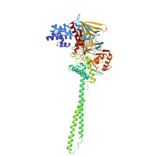
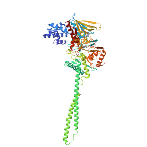
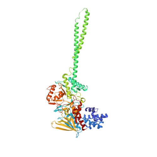
2Z3Y

Crystal structure of Lysine-specific demethylase1


Experimental Data Snapshot

  • Method: X-RAY DIFFRACTION
  • Resolution: 2.25 Å
  • R-Value Free: 0.255 
  • R-Value Work: 0.218 
  • R-Value Observed: 0.218 

wwPDB Validation   3D Report Full Report


Ligand Structure Quality Assessment 


This is version 1.2 of the entry. See complete history


Literature

Crystal structure of histone demethylase LSD1 and tranylcypromine at 2.25A

Mimasu, S.Sengoku, T.Fukuzawa, S.Umehara, T.Yokoyama, S.

(2008) Biochem Biophys Res Commun 366: 15-22

  • DOI: https://doi.org/10.1016/j.bbrc.2007.11.066
  • Primary Citation of Related Structures:  
    2DW4, 2EJR, 2Z3Y, 2Z5U

  • PubMed Abstract: 

    Transcriptional activity and chromatin structure accessibility are correlated with the methylation of specific histone residues. Lysine-specific demethylase 1 (LSD1) is the first discovered histone demethylase, which demethylates Lys4 or Lys9 of histone H3, using FAD. Among the known monoamine oxidase inhibitors, tranylcypromine (Parnate) showed the most potent inhibitory effect on LSD1. Recently, the crystal structure of LSD1 and tranylcypromine was solved at 2.75 A, revealing a five-membered ring fused to the flavin of LSD1. In this study, we refined the crystal structure of the LSD1-tranylcypromine complex to 2.25 A. The five-membered ring model did not fit completely with the electron density, giving R(work)/R(free) values of 0.226/0.254. On the other hand, the N(5) adduct gave the lowest R(work)/R(free) values of 0.218/0.248, among the tested models. These results imply that the LSD1-tranylcypromine complex is not completely composed of the five-membered adduct, but partially contains an intermediate, such as the N(5) adduct.


  • Organizational Affiliation

    Department of Biophysics and Biochemistry, Graduate School of Science, The University of Tokyo, 7-3-1 Hongo, Bunkyo-ku, Tokyo 113-0033, Japan.


Macromolecules
Find similar proteins by:  (by identity cutoff)  |  3D Structure
Entity ID: 1
MoleculeChains Sequence LengthOrganismDetailsImage
Lysine-specific histone demethylase 1662Homo sapiensMutation(s): 0 
Gene Names: KIAA0601LSD1
EC: 1 (PDB Primary Data), 1.14.99.66 (UniProt)
UniProt & NIH Common Fund Data Resources
Find proteins for O60341 (Homo sapiens)
Explore O60341 
Go to UniProtKB:  O60341
PHAROS:  O60341
GTEx:  ENSG00000004487 
Entity Groups  
Sequence Clusters30% Identity50% Identity70% Identity90% Identity95% Identity100% Identity
UniProt GroupO60341
Sequence Annotations
Expand
  • Reference Sequence
Small Molecules
Ligands 1 Unique
IDChains Name / Formula / InChI Key2D Diagram3D Interactions
F2N
Query on F2N

Download Ideal Coordinates CCD File 
B [auth A][(2R,3S,4R,5R)-5-(6-AMINO-9H-PURIN-9-YL)-3,4-DIHYDROXYTETRAHYDROFURAN-2-YL]METHYL (2R,3S,4S)-5-[7,8-DIMETHYL-2,4-DIOXO-5-(3-PHENYLPROPANOYL)-1,3,4,5-TETRAHYDROBENZO[G]PTERIDIN-10(2H)-YL]-2,3,4-TRIHYDROXYPENTYL DIHYDROGEN DIPHOSPHATE
C36 H43 N9 O16 P2
ASOSNULLFLNYFZ-NEHYRMIBSA-N
Experimental Data & Validation

Experimental Data

  • Method: X-RAY DIFFRACTION
  • Resolution: 2.25 Å
  • R-Value Free: 0.255 
  • R-Value Work: 0.218 
  • R-Value Observed: 0.218 
  • Space Group: P 61 2 2
Unit Cell:
Length ( Å )Angle ( ˚ )
a = 185.742α = 90
b = 185.742β = 90
c = 108.725γ = 120
Software Package:
Software NamePurpose
CNSrefinement
HKL-2000data collection
DENZOdata reduction
HKL-2000data scaling
CNSphasing

Structure Validation

View Full Validation Report


Ligand Structure Quality Assessment 


Entry History 

Deposition Data

Revision History  (Full details and data files)

  • Version 1.0: 2008-01-01
    Type: Initial release
  • Version 1.1: 2011-07-13
    Changes: Version format compliance
  • Version 1.2: 2024-03-13
    Changes: Data collection, Database references, Derived calculations