2Z3O | pdb_00002z3o

complex structure of LF-transferase and phenylalanine


Experimental Data Snapshot

  • Method: X-RAY DIFFRACTION
  • Resolution: 2.40 Å
  • R-Value Free: 
    0.269 (Depositor), 0.269 (DCC) 
  • R-Value Work: 
    0.241 (Depositor), 0.240 (DCC) 
  • R-Value Observed: 
    0.241 (Depositor) 

Starting Model: experimental
View more details

wwPDB Validation 3D Report Full Report

Validation slider image for 2Z3O

Ligand Structure Quality Assessment 


This is version 1.2 of the entry. See complete history

Literature

Protein-based peptide-bond formation by aminoacyl-tRNA protein transferase

Watanabe, K.Toh, Y.Suto, K.Shimizu, Y.Oka, N.Wada, T.Tomita, K.

(2007) Nature 449: 867-871

  • DOI: https://doi.org/10.1038/nature06167
  • Primary Citation Related Structures: 
    2Z3K, 2Z3L, 2Z3M, 2Z3N, 2Z3O, 2Z3P

  • PubMed Abstract: 

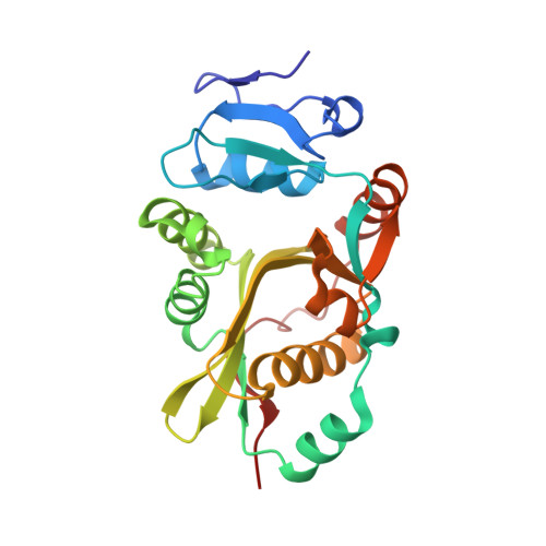
    Eubacterial leucyl/phenylalanyl-tRNA protein transferase (LF-transferase) catalyses peptide-bond formation by using Leu-tRNA(Leu) (or Phe-tRNA(Phe)) and an amino-terminal Arg (or Lys) of a protein, as donor and acceptor substrates, respectively. However, the catalytic mechanism of peptide-bond formation by LF-transferase remained obscure. Here we determine the structures of complexes of LF-transferase and phenylalanyl adenosine, with and without a short peptide bearing an N-terminal Arg. Combining the two separate structures into one structure as well as mutation studies reveal the mechanism for peptide-bond formation by LF-transferase. The electron relay from Asp 186 to Gln 188 helps Gln 188 to attract a proton from the alpha-amino group of the N-terminal Arg of the acceptor peptide. This generates the attacking nucleophile for the carbonyl carbon of the aminoacyl bond of the aminoacyl-tRNA, thus facilitating peptide-bond formation. The protein-based mechanism for peptide-bond formation by LF-transferase is similar to the reverse reaction of the acylation step observed in the peptide hydrolysis reaction by serine proteases.


  • Organizational Affiliation
    • Institute of Biological Resources and Functions, National Institute of Advanced Industrial Sciences and Technology, 1-1-1, Higashi, Tsukuba-shi, Ibaraki 305-8566, Japan.

Macromolecule Content 

  • Total Structure Weight: 53.67 kDa 
  • Atom Count: 3,762 
  • Modeled Residue Count: 463 
  • Deposited Residue Count: 466 
  • Unique protein chains: 1

Macromolecules

Find similar proteins by:|  3D Structure
Entity ID: 1
MoleculeChains  Sequence LengthOrganismDetailsImage
Leucyl/phenylalanyl-tRNA-protein transferase
A, B
233Escherichia coliMutation(s): 0 
Gene Names: Aat
EC: 2.3.2.6
UniProt
Find proteins for P0A8P1 (Escherichia coli (strain K12))
Explore P0A8P1 
Go to UniProtKB:  P0A8P1
Entity Groups
Sequence Clusters30% Identity50% Identity70% Identity90% Identity95% Identity100% Identity
UniProt GroupP0A8P1
Sequence Annotations
Expand
Reference Sequence

Experimental Data & Validation

Experimental Data

  • Method: X-RAY DIFFRACTION
  • Resolution: 2.40 Å
  • R-Value Free:  0.269 (Depositor), 0.269 (DCC) 
  • R-Value Work:  0.241 (Depositor), 0.240 (DCC) 
  • R-Value Observed: 0.241 (Depositor) 
Space Group: P 21 21 2
Unit Cell:
Length ( Å )Angle ( ˚ )
a = 115.49α = 90
b = 129.249β = 90
c = 38.678γ = 90
Software Package:
Software NamePurpose
CNSrefinement
HKL-2000data scaling
CNSphasing

Structure Validation

View Full Validation Report


Ligand Structure Quality Assessment 


Entry History 

Deposition Data

Revision History  (Full details and data files)

  • Version 1.0: 2007-10-23
    Type: Initial release
  • Version 1.1: 2011-07-13
    Changes: Version format compliance
  • Version 1.2: 2023-11-01
    Changes: Data collection, Database references, Refinement description