2Z1J | pdb_00002z1j

Crystal structure of E.coli RNase HI surface charged mutant(Q4R/T40E/Q72H/Q76K/Q80E/T92K/Q105K/Q113R/Q115K/N143K/T145K)


Experimental Data Snapshot

  • Method: X-RAY DIFFRACTION
  • Resolution: 2.38 Å
  • R-Value Free: 
    0.238 (Depositor) 
  • R-Value Work: 
    0.208 (Depositor), 0.203 (DCC) 
  • R-Value Observed: 
    0.208 (Depositor) 

Starting Model: experimental
View more details

wwPDB Validation 3D Report Full Report

Validation slider image for 2Z1J

This is version 1.3 of the entry. See complete history

Literature

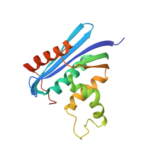
Protein Thermostabilization Requires a Fine-tuned Placement of Surface-charged Residues

You, D.-J.Fukuchi, S.Nishikawa, K.Koga, Y.Takano, K.Kanaya, S.

(2007) J Biochem 142: 507-516

  • DOI: https://doi.org/10.1093/jb/mvm157
  • Primary Citation Related Structures: 
    2Z1G, 2Z1H, 2Z1I, 2Z1J

  • PubMed Abstract: 

    Using the information from the genome projects, recent comparative studies of thermostable proteins have revealed a certain trend of amino acid composition in which polar residues are scarce and charged residues are rich on the protein surface. To clarify experimentally the effect of the amino acid composition of surface residues on the thermostability of Escherichia coli Ribonuclease HI (RNase HI), we constructed six variants in which five to eleven polar residues were replaced by charged residues (5C, 7Ca, 7Cb, 9Ca, 9Cb and 11C). The thermal denaturation experiments indicated that all of the variant proteins are 3.2-10.1 degrees C in Tm less stable than the wild proteins. The crystal structures of resultant protein variants 7Ca, 7Cb, 9Ca and 11C closely resemble that of E. coli RNase HI in their global fold, and several different hydrogen bonding and ion-pair interactions are formed by the mutations. Comparison of the crystal structures of these variant proteins with that of E. coli RNase HI reveals that thermal destabilization is apparently related to electrostatic repulsion of the charged residues with neighbours. This result suggests that charged residues of natural thermostable proteins are strictly posted on the surface with optimal interactions and without repulsive interactions.


  • Organizational Affiliation
    • Department of Material and Life Science, Graduate School of Engineering, Osaka University, 2-1 Yamadaoka, Suita, Osaka 565-0871, Japan.

Macromolecule Content 

  • Total Structure Weight: 17.79 kDa 
  • Atom Count: 1,260 
  • Modeled Residue Count: 151 
  • Deposited Residue Count: 155 
  • Unique protein chains: 1

Macromolecules

Find similar proteins by:|  3D Structure
Entity ID: 1
MoleculeChains  Sequence LengthOrganismDetailsImage
Ribonuclease HI155Escherichia coliMutation(s): 11 
EC: 3.1.26.4
UniProt
Find proteins for P0A7Y4 (Escherichia coli (strain K12))
Explore P0A7Y4 
Go to UniProtKB:  P0A7Y4
Entity Groups
Sequence Clusters30% Identity50% Identity70% Identity90% Identity95% Identity100% Identity
UniProt GroupP0A7Y4
Sequence Annotations
Expand
Reference Sequence

Experimental Data & Validation

Experimental Data

  • Method: X-RAY DIFFRACTION
  • Resolution: 2.38 Å
  • R-Value Free:  0.238 (Depositor) 
  • R-Value Work:  0.208 (Depositor), 0.203 (DCC) 
  • R-Value Observed: 0.208 (Depositor) 
Space Group: P 32 2 1
Unit Cell:
Length ( Å )Angle ( ˚ )
a = 85.273α = 90
b = 85.273β = 90
c = 51.024γ = 120
Software Package:
Software NamePurpose
CNSrefinement
HKL-2000data collection
HKL-2000data reduction
HKL-2000data scaling
AMoREphasing

Structure Validation

View Full Validation Report


Entry History 

Deposition Data

Revision History  (Full details and data files)

  • Version 1.0: 2007-11-13
    Type: Initial release
  • Version 1.1: 2011-07-13
    Changes: Version format compliance
  • Version 1.2: 2021-11-10
    Changes: Database references
  • Version 1.3: 2023-11-01
    Changes: Data collection, Refinement description