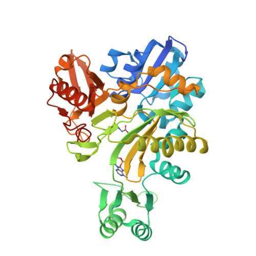
2YS6

Crystal structure of GAR synthetase from Geobacillus kaustophilus


Experimental Data Snapshot

  • Method: X-RAY DIFFRACTION
  • Resolution: 2.21 Å
  • R-Value Free: 0.230 
  • R-Value Work: 0.204 
  • R-Value Observed: 0.204 

Starting Model: experimental
View more details

wwPDB Validation   3D Report Full Report


Ligand Structure Quality Assessment 


This is version 1.2 of the entry. See complete history


Literature

Crystal structures of glycinamide ribonucleotide synthetase, PurD, from thermophilic eubacteria

Sampei, G.Baba, S.Kanagawa, M.Yanai, H.Ishii, T.Kawai, H.Fukai, Y.Ebihara, A.Nakagawa, N.Kawai, G.

(2010) J Biochem 148: 429-438

  • DOI: https://doi.org/10.1093/jb/mvq088
  • Primary Citation of Related Structures:  
    2IP4, 2YRW, 2YRX, 2YS6, 2YS7, 2YW2, 2YYA

  • PubMed Abstract: 

    Glycinamide ribonucleotide synthetase (GAR-syn, PurD) catalyses the second reaction of the purine biosynthetic pathway; the conversion of phosphoribosylamine, glycine and ATP to glycinamide ribonucleotide (GAR), ADP and Pi. In the present study, crystal structures of GAR-syn's from Thermus thermophilus, Geobacillus kaustophilus and Aquifex aeolicus were determined in apo forms. Crystal structures in ligand-bound forms were also determined for G. kaustophilus and A. aeolicus proteins. In general, overall structures of GAR-syn's are similar to each other. However, the orientations of the B domains are varied among GAR-syn's and the MD simulation suggested the mobility of the B domain. Furthermore, it was demonstrated that the B loop in the B domain fixes the position of the β- and γ- phosphate groups of the bound ATP. The structures of GAR-syn's and the bound ligands were compared with each other in detail, and structures of GAR-syn's with full ligands, as well as the possible reaction mechanism, were proposed.


  • Organizational Affiliation

    Department of Applied Physics and Chemistry, Faculty of Electro-Communications, The University of Electro-Communications, 1-5-1 Chofugaoka, Chofu-shi, Tokyo, Japan. sampei@pc.uec.ac.jp


Macromolecules
Find similar proteins by:  (by identity cutoff)  |  3D Structure
Entity ID: 1
MoleculeChains Sequence LengthOrganismDetailsImage
Phosphoribosylglycinamide synthetase451Geobacillus kaustophilusMutation(s): 0 
EC: 6.3.4.13
UniProt
Find proteins for Q5L3C7 (Geobacillus kaustophilus (strain HTA426))
Explore Q5L3C7 
Go to UniProtKB:  Q5L3C7
Entity Groups  
Sequence Clusters30% Identity50% Identity70% Identity90% Identity95% Identity100% Identity
UniProt GroupQ5L3C7
Sequence Annotations
Expand
  • Reference Sequence
Small Molecules
Ligands 2 Unique
IDChains Name / Formula / InChI Key2D Diagram3D Interactions
AMP
Query on AMP

Download Ideal Coordinates CCD File 
C [auth A]ADENOSINE MONOPHOSPHATE
C10 H14 N5 O7 P
UDMBCSSLTHHNCD-KQYNXXCUSA-N
GLY
Query on GLY

Download Ideal Coordinates CCD File 
B [auth A]GLYCINE
C2 H5 N O2
DHMQDGOQFOQNFH-UHFFFAOYSA-N
Experimental Data & Validation

Experimental Data

  • Method: X-RAY DIFFRACTION
  • Resolution: 2.21 Å
  • R-Value Free: 0.230 
  • R-Value Work: 0.204 
  • R-Value Observed: 0.204 
  • Space Group: P 21 21 21
Unit Cell:
Length ( Å )Angle ( ˚ )
a = 50.786α = 90
b = 83.43β = 90
c = 96.536γ = 90
Software Package:
Software NamePurpose
CNSrefinement
HKL-2000data collection
HKL-2000data reduction
HKL-2000data scaling
MOLREPphasing

Structure Validation

View Full Validation Report


Ligand Structure Quality Assessment 


Entry History 

Revision History  (Full details and data files)

  • Version 1.0: 2007-10-09
    Type: Initial release
  • Version 1.1: 2011-07-13
    Changes: Version format compliance
  • Version 1.2: 2023-10-25
    Changes: Data collection, Database references, Derived calculations, Refinement description