2YN2 | pdb_00002yn2

Huf protein - paralogue of the tau55 histidine phosphatase domain


Experimental Data Snapshot

  • Method: X-RAY DIFFRACTION
  • Resolution: 2.05 Å
  • R-Value Free: 
    0.236 (Depositor), 0.256 (DCC) 
  • R-Value Work: 
    0.200 (Depositor) 
  • R-Value Observed: 
    0.202 (Depositor) 

Starting Model: experimental
View more details

wwPDB Validation 3D Report Full Report

Validation slider image for 2YN2

This is version 2.0 of the entry. See complete history

Literature

Structural and Functional Characterization of a Phosphatase Domain within Yeast General Transcription Factor Tfiiic.

Taylor, N.M.I.Glatt, S.Hennrich, M.L.Von Scheven, G.Grotsch, H.Fernandez-Tornero, C.Rybin, V.Gavin, A.Kolb, P.Muller, C.W.

(2013) J Biological Chem 288: 15110

  • DOI: https://doi.org/10.1074/jbc.M112.427856
  • Primary Citation Related Structures: 
    2YN0, 2YN2

  • PubMed Abstract: 

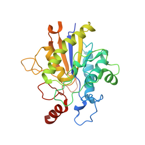
    Saccharomyces cerevisiae τ55, a subunit of the RNA polymerase III-specific general transcription factor TFIIIC, comprises an N-terminal histidine phosphatase domain (τ55-HPD) whose catalytic activity and cellular function is poorly understood. We solved the crystal structures of τ55-HPD and its closely related paralogue Huf and used in silico docking methods to identify phosphoserine- and phosphotyrosine-containing peptides as possible substrates that were subsequently validated using in vitro phosphatase assays. A comparative phosphoproteomic study identified additional phosphopeptides as possible targets that show the involvement of these two phosphatases in the regulation of a variety of cellular functions. Our results identify τ55-HPD and Huf as bona fide protein phosphatases, characterize their substrate specificities, and provide a small set of regulated phosphosite targets in vivo.


  • Organizational Affiliation
    • Structural and Computational Biology Unit, European Molecular Biology Laboratory, 69117 Heidelberg, Germany.

Macromolecule Content 

  • Total Structure Weight: 32.02 kDa 
  • Atom Count: 2,156 
  • Modeled Residue Count: 254 
  • Deposited Residue Count: 279 
  • Unique protein chains: 1

Macromolecules

Find similar proteins by:|  3D Structure
Entity ID: 1
MoleculeChains  Sequence LengthOrganismDetailsImage
UNCHARACTERIZED PROTEIN YNL108C279Saccharomyces cerevisiae S288CMutation(s): 0 
UniProt
Find proteins for P53929 (Saccharomyces cerevisiae (strain ATCC 204508 / S288c))
Explore P53929 
Go to UniProtKB:  P53929
Entity Groups
Sequence Clusters30% Identity50% Identity70% Identity90% Identity95% Identity100% Identity
UniProt GroupP53929
Sequence Annotations
Expand
Reference Sequence

Small Molecules

Ligands 1 Unique
IDChains Name / Formula / InChI Key2D Diagram3D Interactions
FMT

Query on FMT


Download:Ideal Coordinates CCD File
B [auth A]FORMIC ACID
C H2 O2
BDAGIHXWWSANSR-UHFFFAOYSA-N

Experimental Data & Validation

Experimental Data

  • Method: X-RAY DIFFRACTION
  • Resolution: 2.05 Å
  • R-Value Free:  0.236 (Depositor), 0.256 (DCC) 
  • R-Value Work:  0.200 (Depositor) 
  • R-Value Observed: 0.202 (Depositor) 
Space Group: P 32 2 1
Unit Cell:
Length ( Å )Angle ( ˚ )
a = 86.72α = 90
b = 86.72β = 90
c = 98.96γ = 120
Software Package:
Software NamePurpose
PHENIXrefinement
XDSdata reduction
XSCALEdata scaling
PHASERphasing

Structure Validation

View Full Validation Report


Entry History 

Deposition Data

Revision History  (Full details and data files)

  • Version 1.0: 2013-04-03
    Type: Initial release
  • Version 1.1: 2013-04-17
    Changes: Database references
  • Version 1.2: 2013-04-24
    Changes: Structure summary
  • Version 1.3: 2013-06-12
    Changes: Database references
  • Version 2.0: 2023-12-20
    Changes: Atomic model, Data collection, Database references, Derived calculations, Other, Refinement description