2YJL | pdb_00002yjl

Structural characterization of a secretin pilot protein from the type III secretion system (T3SS) of Pseudomonas aeruginosa


Experimental Data Snapshot

  • Method: X-RAY DIFFRACTION
  • Resolution: 1.81 Å
  • R-Value Free: 
    0.262 (Depositor), 0.271 (DCC) 
  • R-Value Work: 
    0.217 (Depositor), 0.222 (DCC) 
  • R-Value Observed: 
    0.221 (Depositor) 

wwPDB Validation 3D Report Full Report

Validation slider image for 2YJL

This is version 1.2 of the entry. See complete history

Literature

Structural Characterization and Membrane Localization of Exsb from the Type III Secretion System (T3Ss) of Pseudomonas Aeruginosa

Izore, T.Perdu, C.Job, V.Atree, I.Faudry, E.Dessen, A.

(2011) J Mol Biology 413: 236

  • DOI: https://doi.org/10.1016/j.jmb.2011.07.043
  • Primary Citation Related Structures: 
    2YJL

  • PubMed Abstract: 

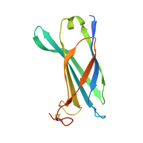
    Pseudomonas aeruginosa is an opportunistic human pathogen that employs a finely tuned type III secretion system (T3SS) to inject toxins directly into the cytoplasm of target cells. ExsB is a 15.6-kDa protein encoded in a T3SS transcription regulation operon that displays high sequence similarity to YscW, a lipoprotein from Yersinia spp. whose genetic neighborhood also involves a transcriptional regulator, and has been shown to play a role in the stabilization of the outer membrane ring of the T3SS. Here, we show that ExsB is expressed in P. aeruginosa upon induction of the T3SS, and subcellular fractionation studies reveal that it is associated with the outer membrane. The high-resolution crystal structure of ExsB shows that it displays a compact β-sandwich fold with interdependent β-sheets. ExsB possesses a large patch of basic residues that could play a role in membrane recognition, and its structure is distinct from that of MxiM, a lipoprotein involved in secretin stabilization in Shigella, as well as from those of Pil lipoproteins involved in pilus biogenesis. These results reveal that small lipoproteins involved in formation of the outer membrane secretin ring display clear structural differences that may be related to the different functions they play in these systems.


  • Organizational Affiliation
    • Bacterial Pathogenesis Group, Institut de Biologie Structurale (IBS), Université Grenoble I, France.

Macromolecule Content 

  • Total Structure Weight: 41.9 kDa 
  • Atom Count: 2,879 
  • Modeled Residue Count: 327 
  • Deposited Residue Count: 375 
  • Unique protein chains: 1

Macromolecules

Find similar proteins by:|  3D Structure
Entity ID: 1
MoleculeChains  Sequence LengthOrganismDetailsImage
EXOENZYME S SYNTHESIS PROTEIN B
A, B, C
125Pseudomonas aeruginosa PAO1Mutation(s): 0 
UniProt
Find proteins for P26994 (Pseudomonas aeruginosa (strain ATCC 15692 / DSM 22644 / CIP 104116 / JCM 14847 / LMG 12228 / 1C / PRS 101 / PAO1))
Explore P26994 
Go to UniProtKB:  P26994
Entity Groups
Sequence Clusters30% Identity50% Identity70% Identity90% Identity95% Identity100% Identity
UniProt GroupP26994
Sequence Annotations
Expand
Reference Sequence

Experimental Data & Validation

Experimental Data

  • Method: X-RAY DIFFRACTION
  • Resolution: 1.81 Å
  • R-Value Free:  0.262 (Depositor), 0.271 (DCC) 
  • R-Value Work:  0.217 (Depositor), 0.222 (DCC) 
  • R-Value Observed: 0.221 (Depositor) 
Space Group: C 1 2 1
Unit Cell:
Length ( Å )Angle ( ˚ )
a = 124.155α = 90
b = 48.358β = 96.92
c = 71.602γ = 90
Software Package:
Software NamePurpose
REFMACrefinement
XDSdata reduction
XSCALEdata scaling
PHENIXphasing

Structure Validation

View Full Validation Report


Entry History 

Deposition Data

Revision History  (Full details and data files)

  • Version 1.0: 2011-08-10
    Type: Initial release
  • Version 1.1: 2011-10-12
    Changes: Database references
  • Version 1.2: 2024-10-16
    Changes: Data collection, Database references, Derived calculations, Other, Structure summary