2YIQ | pdb_00002yiq

Structural analysis of checkpoint kinase 2 in complex with inhibitor PV1322


Experimental Data Snapshot

  • Method: X-RAY DIFFRACTION
  • Resolution: 1.89 Å
  • R-Value Free: 
    0.234 (Depositor), 0.260 (DCC) 
  • R-Value Work: 
    0.197 (Depositor), 0.230 (DCC) 
  • R-Value Observed: 
    0.199 (Depositor) 

Starting Model: experimental
View more details

wwPDB Validation   3D Report Full Report


Ligand Structure Quality Assessment 

Created with Raphaël 2.3.0Worse 01 BetterLigand structure goodness of fit to experimental dataBest fitted YIQClick on this verticalbar to view details

This is version 1.2 of the entry. See complete history


Literature

X-Ray Structures of Checkpoint Kinase 2 in Complex with Inhibitors that Target its Gatekeeper-Dependent Hydrophobic Pocket.

Lountos, G.T.Jobson, A.G.Tropea, J.E.Self, C.R.Zhang, G.Pommier, Y.Shoemaker, R.H.Waugh, D.S.

(2011) FEBS Lett 585: 3245

  • DOI: https://doi.org/10.1016/j.febslet.2011.08.050
  • Primary Citation of Related Structures:  
    2YIQ, 2YIR, 2YIT

  • PubMed Abstract: 

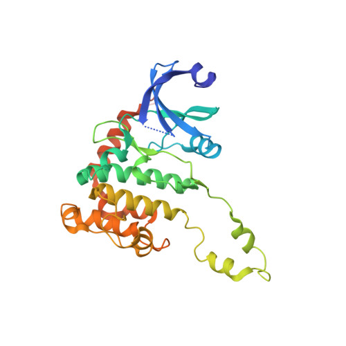
    The serine/threonine checkpoint kinase 2 (Chk2) is an attractive molecular target for the development of small molecule inhibitors to treat cancer. Here, we report the rational design of Chk2 inhibitors that target the gatekeeper-dependent hydrophobic pocket located behind the adenine-binding region of the ATP-binding site. These compounds exhibit IC(50) values in the low nanomolar range and are highly selective for Chk2 over Chk1. X-ray crystallography was used to determine the structures of the inhibitors in complex with the catalytic kinase domain of Chk2 to verify their modes of binding.


  • Organizational Affiliation

    Basic Science Program, SAIC-Frederick, Frederick, MD 21702-1201, USA.


Macromolecules
Find similar proteins by:  (by identity cutoff)  |  3D Structure
Entity ID: 1
MoleculeChains Sequence LengthOrganismDetailsImage
SERINE/THREONINE-PROTEIN KINASE CHK2323Homo sapiensMutation(s): 0 
EC: 2.7.11.1
UniProt & NIH Common Fund Data Resources
Find proteins for O96017 (Homo sapiens)
Explore O96017 
Go to UniProtKB:  O96017
PHAROS:  O96017
GTEx:  ENSG00000183765 
Entity Groups  
Sequence Clusters30% Identity50% Identity70% Identity90% Identity95% Identity100% Identity
UniProt GroupO96017
Sequence Annotations
Expand
  • Reference Sequence
Small Molecules
Ligands 2 Unique
IDChains Name / Formula / InChI Key2D Diagram3D Interactions
YIQ
Query on YIQ

Download Ideal Coordinates CCD File 
B [auth A](E)-5-(1-(2-CARBAMIMIDOYLHYDRAZONO)ETHYL)-N-(1H-INDOL-6-YL)-1H-INDOLE-2-CARBOXAMIDE
C20 H19 N7 O
STGJVWUQAFPAMH-KBKYJPHKSA-N
NO3
Query on NO3

Download Ideal Coordinates CCD File 
C [auth A]NITRATE ION
N O3
NHNBFGGVMKEFGY-UHFFFAOYSA-N
Experimental Data & Validation

Experimental Data

  • Method: X-RAY DIFFRACTION
  • Resolution: 1.89 Å
  • R-Value Free:  0.234 (Depositor), 0.260 (DCC) 
  • R-Value Work:  0.197 (Depositor), 0.230 (DCC) 
  • R-Value Observed: 0.199 (Depositor) 
Space Group: P 32 2 1
Unit Cell:
Length ( Å )Angle ( ˚ )
a = 90.61α = 90
b = 90.61β = 90
c = 93.555γ = 120
Software Package:
Software NamePurpose
REFMACrefinement
HKL-3000data reduction
SCALEPACKdata scaling
MOLREPphasing

Structure Validation

View Full Validation Report


Ligand Structure Quality Assessment 

Created with Raphaël 2.3.0Worse 01 BetterLigand structure goodness of fit to experimental dataBest fitted YIQClick on this verticalbar to view details

Entry History 

Deposition Data

Revision History  (Full details and data files)

  • Version 1.0: 2011-09-07
    Type: Initial release
  • Version 1.1: 2011-10-26
    Changes: Database references
  • Version 1.2: 2023-12-20
    Changes: Data collection, Database references, Derived calculations, Other, Refinement description