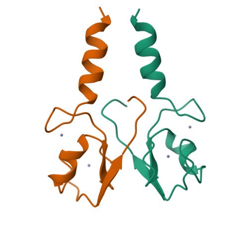
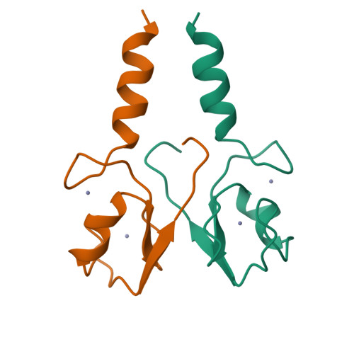
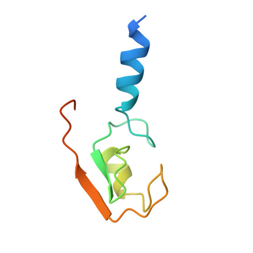
2YHN

The IDOL-UBE2D complex mediates sterol-dependent degradation of the LDL receptor


Experimental Data Snapshot

  • Method: X-RAY DIFFRACTION
  • Resolution: 3.00 Å
  • R-Value Free: 0.297 
  • R-Value Work: 0.228 
  • R-Value Observed: 0.232 

Starting Model: experimental
View more details

wwPDB Validation   3D Report Full Report


This is version 1.2 of the entry. See complete history


Literature

The Idol-Ube2D Complex Mediates Sterol-Dependent Degradation of the Ldl Receptor.

Zhang, L.Fairall, L.Goult, B.T.Calkin, A.C.Hong, C.Millard, C.J.Tontonoz, P.Schwabe, J.W.R.

(2011) Genes Dev 25: 1262

  • DOI: https://doi.org/10.1101/gad.2056211
  • Primary Citation of Related Structures:  
    2YHN, 2YHO

  • PubMed Abstract: 

    We previously identified the E3 ubiquitin ligase IDOL as a sterol-dependent regulator of the LDL receptor (LDLR). The molecular pathway underlying IDOL action, however, remains to be determined. Here we report the identification and biochemical and structural characterization of an E2-E3 ubiquitin ligase complex for LDLR degradation. We identified the UBE2D family (UBE2D1-4) as E2 partners for IDOL that support both autoubiquitination and IDOL-dependent ubiquitination of the LDLR in a cell-free system. NMR chemical shift mapping and a 2.1 Å crystal structure of the IDOL RING domain-UBE2D1 complex revealed key interactions between the dimeric IDOL protein and the E2 enzyme. Analysis of the IDOL-UBE2D1 interface also defined the stereochemical basis for the selectivity of IDOL for UBE2Ds over other E2 ligases. Structure-based mutations that inhibit IDOL dimerization or IDOL-UBE2D interaction block IDOL-dependent LDLR ubiquitination and degradation. Furthermore, expression of a dominant-negative UBE2D enzyme inhibits the ability of IDOL to degrade the LDLR in cells. These results identify the IDOL-UBE2D complex as an important determinant of LDLR activity, and provide insight into molecular mechanisms underlying the regulation of cholesterol uptake.


  • Organizational Affiliation

    Howard Hughes Medical Institute, University of California at Los Angeles School of Medicine, USA.


Macromolecules
Find similar proteins by:  (by identity cutoff)  |  3D Structure
Entity ID: 1
MoleculeChains Sequence LengthOrganismDetailsImage
E3 UBIQUITIN-PROTEIN LIGASE MYLIP
A, B
79Homo sapiensMutation(s): 0 
EC: 6.3.2 (PDB Primary Data), 2.3.2.27 (UniProt)
UniProt & NIH Common Fund Data Resources
Find proteins for Q8WY64 (Homo sapiens)
Explore Q8WY64 
Go to UniProtKB:  Q8WY64
PHAROS:  Q8WY64
GTEx:  ENSG00000007944 
Entity Groups  
Sequence Clusters30% Identity50% Identity70% Identity90% Identity95% Identity100% Identity
UniProt GroupQ8WY64
Sequence Annotations
Expand
  • Reference Sequence
Experimental Data & Validation

Experimental Data

  • Method: X-RAY DIFFRACTION
  • Resolution: 3.00 Å
  • R-Value Free: 0.297 
  • R-Value Work: 0.228 
  • R-Value Observed: 0.232 
  • Space Group: I 1 2 1
Unit Cell:
Length ( Å )Angle ( ˚ )
a = 79.26α = 90
b = 22.77β = 116.82
c = 87.838γ = 90
Software Package:
Software NamePurpose
PHENIXrefinement
MOSFLMdata reduction
SCALAdata scaling
PHASERphasing

Structure Validation

View Full Validation Report


Entry History 

Deposition Data

Revision History  (Full details and data files)

  • Version 1.0: 2011-06-29
    Type: Initial release
  • Version 1.1: 2011-07-13
    Changes: Version format compliance
  • Version 1.2: 2023-12-20
    Changes: Data collection, Database references, Derived calculations, Other, Refinement description