2YFL

Crystal Structure of Biphenyl dioxygenase variant RR41 with 2-chloro dibenzofuran


Experimental Data Snapshot

  • Method: X-RAY DIFFRACTION
  • Resolution: 2.60 Å
  • R-Value Free: 0.280 
  • R-Value Work: 0.215 
  • R-Value Observed: 0.218 

Starting Model: experimental
View more details

wwPDB Validation   3D Report Full Report


Ligand Structure Quality Assessment 


This is version 1.2 of the entry. See complete history


Literature

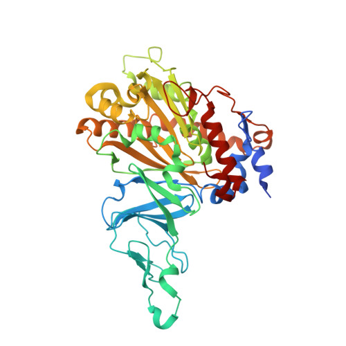
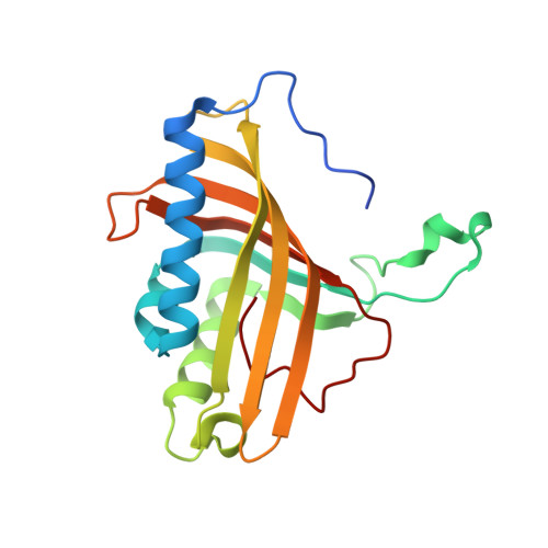
Structural Insights Into the Metabolism of 2-Chlorodibenzofuran by an Evolved Biphenyl Dioxygenase.

Kumar, P.Mohammadi, M.Dhindwal, S.Pham, T.T.Bolin, J.T.Sylvestre, M.

(2012) Biochem Biophys Res Commun 421: 757

  • DOI: https://doi.org/10.1016/j.bbrc.2012.04.078
  • Primary Citation of Related Structures:  
    2YFL

  • PubMed Abstract: 

    The biphenyl dioxygenase of Burkholderia xenovorans LB400 (BphAE(LB400)) is a Rieske-type oxygenase that catalyzes the stereospecific oxygenation of many heterocyclic aromatics including dibenzofuran. In a previous work, we evolved BphAE(LB400) and obtained BphAE(RR41). This variant metabolizes dibenzofuran and 2-chlorodibenzofuran more efficiently than BphAE(LB400). However, the regiospecificity of BphAE(RR41) toward these substrates differs. Dibenzofuran is metabolized principally through a lateral dioxygenation whereas 2-chlorodibenzofuran is metabolized principally through an angular dioxygenation. In order to explain this difference, we examined the crystal structures of both substrate-bound forms of BphAE(RR41) obtained under anaerobic conditions. This structure analysis, in combination with biochemical data for a Ser283Gly mutant provided evidences that the substrate is compelled to move after oxygen-binding in BphAE(RR41):dibenzofuran. In BphAE(RR41):2-chlorodibenzofuran, the chlorine atom is close to the side chain of Ser283. This contact is missing in the BphAE(RR41):dibenzofuran, and strong enough in the BphAE(RR41):2-chlorodibenzofuran to help prevent substrate movement during the catalytic reaction.


  • Organizational Affiliation

    Department of Biological Sciences and Center for Cancer Research, Purdue University, West Lafayette, IN 47907, USA.


Macromolecules
Find similar proteins by:  (by identity cutoff)  |  3D Structure
Entity ID: 1
MoleculeChains Sequence LengthOrganismDetailsImage
BIPHENYL DIOXYGENASE SUBUNIT ALPHA
A, C, E, G, I
A, C, E, G, I, K
459Paraburkholderia xenovorans LB400Mutation(s): 5 
EC: 1.14.12.18
UniProt
Find proteins for P37333 (Paraburkholderia xenovorans (strain LB400))
Explore P37333 
Go to UniProtKB:  P37333
Entity Groups  
Sequence Clusters30% Identity50% Identity70% Identity90% Identity95% Identity100% Identity
UniProt GroupP37333
Sequence Annotations
Expand
  • Reference Sequence
Find similar proteins by:  (by identity cutoff)  |  3D Structure
Entity ID: 2
MoleculeChains Sequence LengthOrganismDetailsImage
BIPHENYL DIOXYGENASE SUBUNIT BETA
B, D, F, H, J
B, D, F, H, J, L
188Paraburkholderia xenovorans LB400Mutation(s): 0 
EC: 1.14.12.18
UniProt
Find proteins for P37334 (Paraburkholderia xenovorans (strain LB400))
Explore P37334 
Go to UniProtKB:  P37334
Entity Groups  
Sequence Clusters30% Identity50% Identity70% Identity90% Identity95% Identity100% Identity
UniProt GroupP37334
Sequence Annotations
Expand
  • Reference Sequence
Small Molecules
Ligands 3 Unique
IDChains Name / Formula / InChI Key2D Diagram3D Interactions
DC4
Query on DC4

Download Ideal Coordinates CCD File 
O [auth A],
R [auth C]
2-CHLORODIBENZOFURAN
C12 H7 Cl O
PRKTYWJFCODJOA-UHFFFAOYSA-N
FES
Query on FES

Download Ideal Coordinates CCD File 
M [auth A]
P [auth C]
S [auth E]
U [auth G]
W [auth I]
M [auth A],
P [auth C],
S [auth E],
U [auth G],
W [auth I],
Y [auth K]
FE2/S2 (INORGANIC) CLUSTER
Fe2 S2
NIXDOXVAJZFRNF-UHFFFAOYSA-N
FE2
Query on FE2

Download Ideal Coordinates CCD File 
N [auth A]
Q [auth C]
T [auth E]
V [auth G]
X [auth I]
N [auth A],
Q [auth C],
T [auth E],
V [auth G],
X [auth I],
Z [auth K]
FE (II) ION
Fe
CWYNVVGOOAEACU-UHFFFAOYSA-N
Experimental Data & Validation

Experimental Data

  • Method: X-RAY DIFFRACTION
  • Resolution: 2.60 Å
  • R-Value Free: 0.280 
  • R-Value Work: 0.215 
  • R-Value Observed: 0.218 
  • Space Group: P 1 21 1
Unit Cell:
Length ( Å )Angle ( ˚ )
a = 86.556α = 90
b = 275.982β = 117.46
c = 92.057γ = 90
Software Package:
Software NamePurpose
REFMACrefinement
HKL-2000data reduction
SCALEPACKdata scaling
MOLREPphasing

Structure Validation

View Full Validation Report


Ligand Structure Quality Assessment 


Entry History 

Deposition Data

Revision History  (Full details and data files)

  • Version 1.0: 2012-05-16
    Type: Initial release
  • Version 1.1: 2012-05-30
    Changes: Other
  • Version 1.2: 2023-12-20
    Changes: Data collection, Database references, Derived calculations, Other, Refinement description