2YEV | pdb_00002yev

Structure of caa3-type cytochrome oxidase


Experimental Data Snapshot

  • Method: X-RAY DIFFRACTION
  • Resolution: 2.36 Å
  • R-Value Free: 
    0.218 (Depositor), 0.215 (DCC) 
  • R-Value Work: 
    0.171 (Depositor), 0.168 (DCC) 
  • R-Value Observed: 
    0.174 (Depositor) 

Starting Model: experimental
View more details

wwPDB Validation 3D Report Full Report

Validation slider image for 2YEV

Ligand Structure Quality Assessment 


This is version 1.9 of the entry. See complete history

Literature

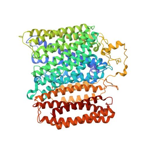
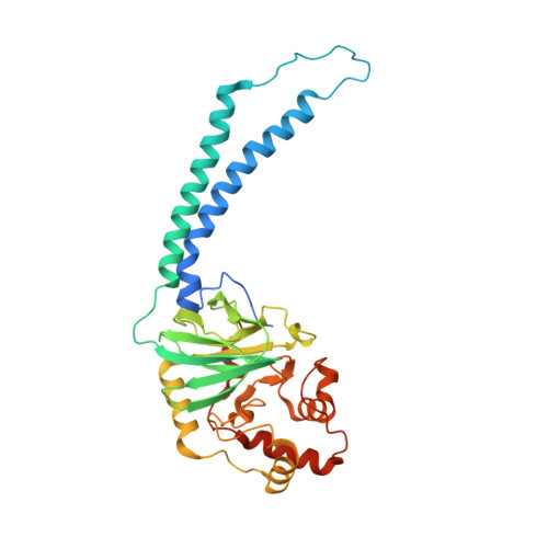
Structural Insights Into Electron Transfer in Caa3-Type Cytochrome Oxidases.

Lyons, J.A.Aragao, D.Slattery, O.Pisliakov, A.V.Soulimane, T.Caffrey, M.

(2012) Nature 487: 514

  • DOI: https://doi.org/10.1038/nature11182
  • Primary Citation Related Structures: 
    2YEV

  • PubMed Abstract: 

    Cytochrome c oxidase is a member of the haem copper oxidase superfamily (HCO). HCOs function as the terminal enzymes in the respiratory chain of mitochondria and aerobic prokaryotes, coupling molecular oxygen reduction to transmembrane proton pumping. Integral to the enzyme's function is the transfer of electrons from cytochrome c to the oxidase via a transient association of the two proteins. Electron entry and exit are proposed to occur from the same site on cytochrome c. Here we report the crystal structure of the caa3-type cytochrome oxidase from Thermus thermophilus, which has a covalently tethered cytochrome c domain. Crystals were grown in a bicontinuous mesophase using a synthetic short-chain monoacylglycerol as the hosting lipid. From the electron density map, at 2.36 Å resolution, a novel integral membrane subunit and a native glycoglycerophospholipid embedded in the complex were identified. Contrary to previous electron transfer mechanisms observed for soluble cytochrome c, the structure reveals the architecture of the electron transfer complex for the fused cupredoxin/cytochrome c domain, which implicates different sites on cytochrome c for electron entry and exit. Support for an alternative to the classical proton gate characteristic of this HCO class is presented.


  • Organizational Affiliation
    • Department of Chemical and Environmental Sciences, University of Limerick, Limerick, Ireland.

Macromolecule Content 

  • Total Structure Weight: 277.44 kDa 
  • Atom Count: 19,587 
  • Modeled Residue Count: 2,325 
  • Deposited Residue Count: 2,388 
  • Unique protein chains: 3

Macromolecules

Find similar proteins by:|  3D Structure
Entity ID: 1
MoleculeChains  Sequence LengthOrganismDetailsImage
CYTOCHROME C OXIDASE POLYPEPTIDE I+III
A, D
791Thermus thermophilus HB8Mutation(s): 0 
EC: 1.9.3.1 (PDB Primary Data), 7.1.1.9 (UniProt)
Membrane Entity: Yes 
UniProt
Find proteins for P98005 (Thermus thermophilus (strain ATCC 27634 / DSM 579 / HB8))
Explore P98005 
Go to UniProtKB:  P98005
Entity Groups
Sequence Clusters30% Identity50% Identity70% Identity90% Identity95% Identity100% Identity
UniProt GroupP98005
Sequence Annotations
Expand
Reference Sequence
Find similar proteins by:|  3D Structure
Entity ID: 2
MoleculeChains  Sequence LengthOrganismDetailsImage
CYTOCHROME C OXIDASE SUBUNIT 2
B, E
337Thermus thermophilus HB8Mutation(s): 0 
EC: 1.9.3.1 (PDB Primary Data), 7.1.1.9 (UniProt)
Membrane Entity: Yes 
UniProt
Find proteins for Q5SLI2 (Thermus thermophilus (strain ATCC 27634 / DSM 579 / HB8))
Explore Q5SLI2 
Go to UniProtKB:  Q5SLI2
Entity Groups
Sequence Clusters30% Identity50% Identity70% Identity90% Identity95% Identity100% Identity
UniProt GroupQ5SLI2
Sequence Annotations
Expand
Reference Sequence
Find similar proteins by:|  3D Structure
Entity ID: 3
MoleculeChains  Sequence LengthOrganismDetailsImage
CAA3-TYPE CYTOCHROME OXIDASE SUBUNIT IV
C, F
66Thermus thermophilus HB8Mutation(s): 0 
EC: 1.9.3.1
Membrane Entity: Yes 
UniProt
Find proteins for Q5SH67 (Thermus thermophilus (strain ATCC 27634 / DSM 579 / HB8))
Explore Q5SH67 
Go to UniProtKB:  Q5SH67
Entity Groups
Sequence Clusters30% Identity50% Identity70% Identity90% Identity95% Identity100% Identity
UniProt GroupQ5SH67
Sequence Annotations
Expand
Reference Sequence

Small Molecules

Ligands 10 Unique
IDChains Name / Formula / InChI Key2D Diagram3D Interactions
5PL

Query on 5PL


Download:Ideal Coordinates CCD File
G [auth A],
S [auth D]
(1R,4S,6R)-6-({[2-(ACETYLAMINO)-2-DEOXY-ALPHA-D-GLUCOPYRANOSYL]OXY}METHYL)-4-HYDROXY-1-{[(15-METHYLHEXADECANOYL)OXY]METHYL}-4-OXIDO-7-OXO-3,5-DIOXA-8-AZA-4-PHOSPHAHEPTACOS-1-YL 15-METHYLHEXADECANOATE
C67 H129 N2 O15 P
LEQHPGOQHCQGAI-TYASGWLASA-N
HAS

Query on HAS


Download:Ideal Coordinates CCD File
H [auth A],
I [auth A],
T [auth D],
U [auth D]
HEME-AS
C54 H64 Fe N4 O6
PDYODZVCODUKFH-ZOMLSHGTSA-L
HEC

Query on HEC


Download:Ideal Coordinates CCD File
P [auth B],
Z [auth E]
HEME C
C34 H34 Fe N4 O4
HXQIYSLZKNYNMH-LJNAALQVSA-N
4AG

Query on 4AG


Download:Ideal Coordinates CCD File
K [auth A](2R)-3-HYDROXYPROPANE-1,2-DIYL DIHEXADECANOATE
C35 H68 O5
JEJLGIQLPYYGEE-MGBGTMOVSA-N
7E9

Query on 7E9


Download:Ideal Coordinates CCD File
Q [auth B]1,3-DIHYDROXYPROPAN-2-YL (Z)-TETRADEC-7-ENOATE
C17 H32 O4
SSMGVWSPWWDBRB-FPLPWBNLSA-N
7E8

Query on 7E8


Download:Ideal Coordinates CCD File
L [auth A],
M [auth A]
(2R)-2,3-DIHYDROXYPROPYL (7Z)-TETRADEC-7-ENOATE
C17 H32 O4
LVBAGTJIDOCNIJ-XITLMJRVSA-N
CUA

Query on CUA


Download:Ideal Coordinates CCD File
O [auth B],
Y [auth E]
DINUCLEAR COPPER ION
Cu2
ALKZAGKDWUSJED-UHFFFAOYSA-N
CU

Query on CU


Download:Ideal Coordinates CCD File
J [auth A],
V [auth D]
COPPER (II) ION
Cu
JPVYNHNXODAKFH-UHFFFAOYSA-N
CL

Query on CL


Download:Ideal Coordinates CCD File
AA [auth F],
R [auth C],
W [auth D]
CHLORIDE ION
Cl
VEXZGXHMUGYJMC-UHFFFAOYSA-M
MG

Query on MG


Download:Ideal Coordinates CCD File
N [auth A],
X [auth D]
MAGNESIUM ION
Mg
JLVVSXFLKOJNIY-UHFFFAOYSA-N
Modified Residues  1 Unique
IDChains TypeFormula2D DiagramParent
FME
Query on FME
C, F
L-PEPTIDE LINKINGC6 H11 N O3 SMET

Experimental Data & Validation

Experimental Data

  • Method: X-RAY DIFFRACTION
  • Resolution: 2.36 Å
  • R-Value Free:  0.218 (Depositor), 0.215 (DCC) 
  • R-Value Work:  0.171 (Depositor), 0.168 (DCC) 
  • R-Value Observed: 0.174 (Depositor) 
Space Group: I 1 2 1
Unit Cell:
Length ( Å )Angle ( ˚ )
a = 127.25α = 90
b = 76.03β = 92.21
c = 300.267γ = 90
Software Package:
Software NamePurpose
PHENIXrefinement
XDSdata reduction
XSCALEdata scaling
PHASERphasing

Structure Validation

View Full Validation Report


Ligand Structure Quality Assessment 


Entry History 

Deposition Data

Revision History  (Full details and data files)

  • Version 1.0: 2012-05-23
    Type: Initial release
  • Version 1.1: 2012-06-27
    Changes: Other
  • Version 1.2: 2012-08-01
    Changes: Database references
  • Version 1.3: 2015-04-29
    Changes: Data collection
  • Version 1.4: 2019-03-06
    Changes: Data collection, Derived calculations, Experimental preparation, Other
  • Version 1.5: 2019-05-08
    Changes: Advisory, Data collection, Derived calculations
  • Version 1.6: 2019-07-10
    Changes: Data collection
  • Version 1.7: 2023-03-29
    Changes: Database references, Derived calculations, Other, Structure summary
  • Version 1.8: 2024-02-07
    Changes: Data collection, Refinement description
  • Version 1.9: 2024-11-13
    Changes: Structure summary