2YDR

CpOGA D298N in complex with p53-derived O-GlcNAc peptide


Experimental Data Snapshot

  • Method: X-RAY DIFFRACTION
  • Resolution: 2.75 Å
  • R-Value Free: 0.219 
  • R-Value Work: 0.185 
  • R-Value Observed: 0.186 

Starting Model: experimental
View more details

wwPDB Validation   3D Report Full Report


Ligand Structure Quality Assessment 


This is version 1.2 of the entry. See complete history


Literature

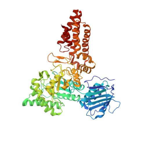
Synergy of Peptide and Sugar in O-Glcnacase Substrate Recognition.

Schimpl, M.Borodkin, V.S.Gray, L.J.Van Aalten, D.M.F.

(2012) Chem Biol 19: 173

  • DOI: https://doi.org/10.1016/j.chembiol.2012.01.011
  • Primary Citation of Related Structures:  
    2YDQ, 2YDR, 2YDS

  • PubMed Abstract: 

    Protein O-GlcNAcylation is an essential reversible posttranslational modification in higher eukaryotes. O-GlcNAc addition and removal is catalyzed by O-GlcNAc transferase and O-GlcNAcase, respectively. We report the molecular details of the interaction of a bacterial O-GlcNAcase homolog with three different synthetic glycopeptides derived from characterized O-GlcNAc sites in the human proteome. Strikingly, the peptides bind a conserved O-GlcNAcase substrate binding groove with similar orientation and conformation. In addition to extensive contacts with the sugar, O-GlcNAcase recognizes the peptide backbone through hydrophobic interactions and intramolecular hydrogen bonds, while avoiding interactions with the glycopeptide side chains. These findings elucidate the molecular basis of O-GlcNAcase substrate specificity, explaining how a single enzyme achieves cycling of the complete O-GlcNAc proteome. In addition, this work will aid development of O-GlcNAcase inhibitors that target the peptide binding site.


  • Organizational Affiliation

    Division of Cell Signalling & Immunology, College of Life Sciences, University of Dundee, Dundee DD1 5EH, Scotland.


Macromolecules
Find similar proteins by:  (by identity cutoff)  |  3D Structure
Entity ID: 1
MoleculeChains Sequence LengthOrganismDetailsImage
O-GLCNACASE NAGJ590Clostridium perfringensMutation(s): 1 
EC: 3.2.1.52
UniProt
Find proteins for Q0TR53 (Clostridium perfringens (strain ATCC 13124 / DSM 756 / JCM 1290 / NCIMB 6125 / NCTC 8237 / Type A))
Explore Q0TR53 
Go to UniProtKB:  Q0TR53
Entity Groups  
Sequence Clusters30% Identity50% Identity70% Identity90% Identity95% Identity100% Identity
UniProt GroupQ0TR53
Sequence Annotations
Expand
  • Reference Sequence

Find similar proteins by:  Sequence   |   3D Structure  

Entity ID: 2
MoleculeChains Sequence LengthOrganismDetailsImage
CELLULAR TUMOR ANTIGEN P53B [auth P]11Homo sapiensMutation(s): 0 
UniProt & NIH Common Fund Data Resources
Find proteins for P04637 (Homo sapiens)
Explore P04637 
Go to UniProtKB:  P04637
GTEx:  ENSG00000141510 
Entity Groups  
Sequence Clusters30% Identity50% Identity70% Identity90% Identity95% Identity100% Identity
UniProt GroupP04637
Glycosylation
Glycosylation Sites: 1Go to GlyGen: P04637-1
Sequence Annotations
Expand
  • Reference Sequence
Small Molecules
Ligands 2 Unique
IDChains Name / Formula / InChI Key2D Diagram3D Interactions
NAG
Query on NAG

Download Ideal Coordinates CCD File 
U [auth P]2-acetamido-2-deoxy-beta-D-glucopyranose
C8 H15 N O6
OVRNDRQMDRJTHS-FMDGEEDCSA-N
CD
Query on CD

Download Ideal Coordinates CCD File 
C [auth A]
D [auth A]
E [auth A]
F [auth A]
G [auth A]
C [auth A],
D [auth A],
E [auth A],
F [auth A],
G [auth A],
H [auth A],
I [auth A],
J [auth A],
K [auth A],
L [auth A],
M [auth A],
N [auth A],
O [auth A],
P [auth A],
Q [auth A],
R [auth A],
S [auth A],
T [auth A]
CADMIUM ION
Cd
WLZRMCYVCSSEQC-UHFFFAOYSA-N
Experimental Data & Validation

Experimental Data

  • Method: X-RAY DIFFRACTION
  • Resolution: 2.75 Å
  • R-Value Free: 0.219 
  • R-Value Work: 0.185 
  • R-Value Observed: 0.186 
  • Space Group: P 61
Unit Cell:
Length ( Å )Angle ( ˚ )
a = 117.95α = 90
b = 117.95β = 90
c = 148.121γ = 120
Software Package:
Software NamePurpose
REFMACrefinement
MOSFLMdata reduction
SCALAdata scaling
MOLREPphasing

Structure Validation

View Full Validation Report


Ligand Structure Quality Assessment 


Entry History 

Deposition Data

Revision History  (Full details and data files)

  • Version 1.0: 2012-03-14
    Type: Initial release
  • Version 1.1: 2020-07-29
    Type: Remediation
    Reason: Carbohydrate remediation
    Changes: Data collection, Derived calculations, Other, Structure summary
  • Version 1.2: 2023-12-20
    Changes: Data collection, Database references, Refinement description, Structure summary