Structurally Sophisticated Octahedral Metal Complexes as Highly Selective Protein Kinase Inhibitors.
Feng, L., Geisselbrecht, Y., Blanck, S., Wilbuer, A., Atilla-Gokcumen, G.E., Filippakopoulos, P., Kraeling, K., Celik, M.A., Harms, K., Maksimoska, J., Marmorstein, R., Frenking, G., Knapp, S., Essen, L.O., Meggers, E.(2011) J Am Chem Soc 133: 5976
- PubMed: 21446733
- DOI: https://doi.org/10.1021/ja1112996
- Primary Citation of Related Structures:
2YAK, 3PUP - PubMed Abstract:
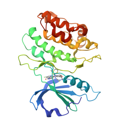
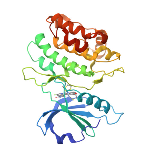
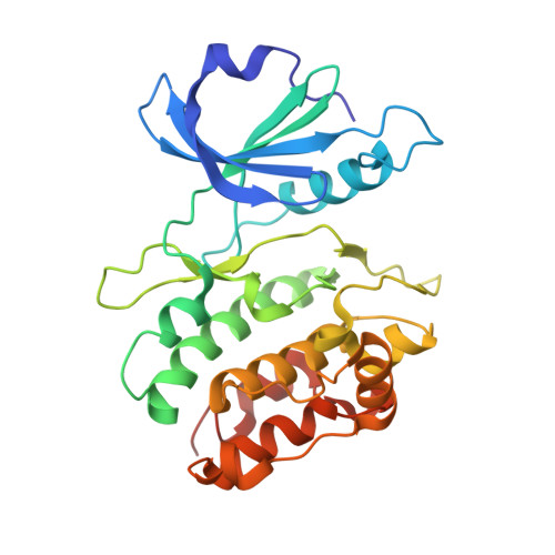
The generation of synthetic compounds with exclusive target specificity is an extraordinary challenge of molecular recognition and demands novel design strategies, in particular for large and homologous protein families such as protein kinases with more than 500 members. Simple organic molecules often do not reach the necessary sophistication to fulfill this task. Here, we present six carefully tailored, stable metal-containing compounds in which unique and defined molecular geometries with natural-product-like structural complexity are constructed around octahedral ruthenium(II) or iridium(III) metal centers. Each of the six reported metal compounds displays high selectivity for an individual protein kinase, namely GSK3α, PAK1, PIM1, DAPK1, MLCK, and FLT4. Although being conventional ATP-competitive inhibitors, the combination of the unusual globular shape and rigidity characteristics, of these compounds facilitates the design of highly selective protein kinase inhibitors. Unique structural features of the octahedral coordination geometry allow novel interactions with the glycine-rich loop, which contribute significantly to binding potencies and selectivities. The sensitive correlation between metal coordination sphere and inhibition properties suggests that in this design, the metal is located at a "hot spot" within the ATP binding pocket, not too close to the hinge region where globular space is unavailable, and at the same time not too far out toward the solvent where the octahedral coordination sphere would not have a significant impact on potency and selectivity. This study thus demonstrates that inert (stable) octahedral metal complexes are sophisticated structural scaffolds for the design of highly selective chemical probes.
Organizational Affiliation:
Fachbereich Chemie, Philipps-Universität Marburg, Hans-Meerwein-Strasse, 35043 Marburg, Germany.


