2Y9W | pdb_00002y9w

Crystal structure of PPO3, a tyrosinase from Agaricus bisporus, in deoxy-form that contains additional unknown lectin-like subunit


Experimental Data Snapshot

  • Method: X-RAY DIFFRACTION
  • Resolution: 2.30 Å
  • R-Value Free: 
    0.237 (Depositor), 0.237 (DCC) 
  • R-Value Work: 
    0.181 (Depositor), 0.182 (DCC) 
  • R-Value Observed: 
    0.184 (Depositor) 

Starting Models: experimental
View more details

wwPDB Validation 3D Report Full Report

Validation slider image for 2Y9W

This is version 1.3 of the entry. See complete history

Literature

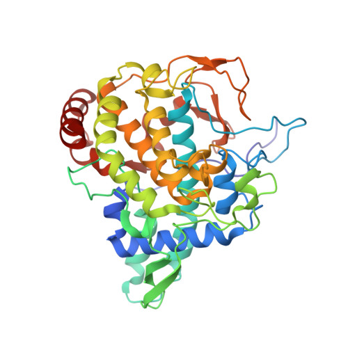
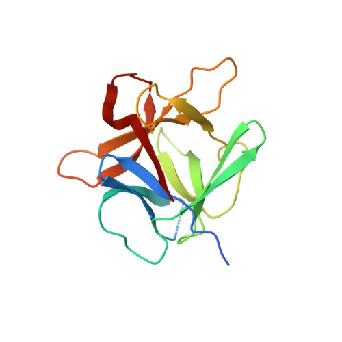
Crystal Structure of Agaricus Bisporus Mushroom Tyrosinase: Identity of the Tetramer Subunits and Interaction with Tropolone.

Ismaya, W.T.Rozeboom, H.J.Weijn, A.Mes, J.J.Fusetti, F.Wichers, H.J.Dijkstra, B.W.

(2011) Biochemistry 50: 5477

  • DOI: https://doi.org/10.1021/bi200395t
  • Primary Citation Related Structures: 
    2Y9W, 2Y9X

  • PubMed Abstract: 

    Tyrosinase catalyzes the conversion of phenolic compounds into their quinone derivatives, which are precursors for the formation of melanin, a ubiquitous pigment in living organisms. Because of its importance for browning reactions in the food industry, the tyrosinase from the mushroom Agaricus bisporus has been investigated in depth. In previous studies the tyrosinase enzyme complex was shown to be a H(2)L(2) tetramer, but no clues were obtained of the identities of the subunits, their mode of association, and the 3D structure of the complex. Here we unravel this tetramer at the molecular level. Its 2.3 Å resolution crystal structure is the first structure of the full fungal tyrosinase complex. The complex comprises two H subunits of ∼392 residues and two L subunits of ∼150 residues. The H subunit originates from the ppo3 gene and has a fold similar to other tyrosinases, but it is ∼100 residues larger. The L subunit appeared to be the product of orf239342 and has a lectin-like fold. The H subunit contains a binuclear copper-binding site in the deoxy-state, in which three histidine residues coordinate each copper ion. The side chains of these histidines have their orientation fixed by hydrogen bonds or, in the case of His85, by a thioether bridge with the side chain of Cys83. The specific tyrosinase inhibitor tropolone forms a pre-Michaelis complex with the enzyme. It binds near the binuclear copper site without directly coordinating the copper ions. The function of the ORF239342 subunits is not known. Carbohydrate binding sites identified in other lectins are not conserved in ORF239342, and the subunits are over 25 Å away from the active site, making a role in activity unlikely. The structures explain how calcium ions stabilize the tetrameric state of the enzyme.


  • Organizational Affiliation
    • Laboratory of Biophysical Chemistry, University of Groningen, Groningen, The Netherlands.

Macromolecule Content 

  • Total Structure Weight: 125.31 kDa 
  • Atom Count: 8,962 
  • Modeled Residue Count: 1,053 
  • Deposited Residue Count: 1,082 
  • Unique protein chains: 2

Macromolecules

Find similar proteins by:|  3D Structure
Entity ID: 1
MoleculeChains  Sequence LengthOrganismDetailsImage
POLYPHENOL OXIDASE
A, B
391Agaricus bisporusMutation(s): 0 
EC: 1.14.18.1
UniProt
Find proteins for C7FF04 (Agaricus bisporus)
Explore C7FF04 
Go to UniProtKB:  C7FF04
Entity Groups
Sequence Clusters30% Identity50% Identity70% Identity90% Identity95% Identity100% Identity
UniProt GroupC7FF04
Sequence Annotations
Expand
Reference Sequence
Find similar proteins by:|  3D Structure
Entity ID: 2
MoleculeChains  Sequence LengthOrganismDetailsImage
LECTIN-LIKE FOLD PROTEIN
C, D
150Agaricus bisporusMutation(s): 0 
UniProt
Find proteins for G1K3P4 (Agaricus bisporus)
Explore G1K3P4 
Go to UniProtKB:  G1K3P4
Entity Groups
Sequence Clusters30% Identity50% Identity70% Identity90% Identity95% Identity100% Identity
UniProt GroupG1K3P4
Sequence Annotations
Expand
Reference Sequence

Small Molecules

Ligands 4 Unique
IDChains Name / Formula / InChI Key2D Diagram3D Interactions
HO

Query on HO


Download:Ideal Coordinates CCD File
G [auth A]
H [auth A]
I [auth A]
M [auth B]
N [auth B]
G [auth A],
H [auth A],
I [auth A],
M [auth B],
N [auth B],
O [auth B]
HOLMIUM ATOM
Ho
KJZYNXUDTRRSPN-UHFFFAOYSA-N
PGE

Query on PGE


Download:Ideal Coordinates CCD File
R [auth D]TRIETHYLENE GLYCOL
C6 H14 O4
ZIBGPFATKBEMQZ-UHFFFAOYSA-N
PEG

Query on PEG


Download:Ideal Coordinates CCD File
J [auth A],
P [auth B],
Q [auth C]
DI(HYDROXYETHYL)ETHER
C4 H10 O3
MTHSVFCYNBDYFN-UHFFFAOYSA-N
CU

Query on CU


Download:Ideal Coordinates CCD File
E [auth A],
F [auth A],
K [auth B],
L [auth B]
COPPER (II) ION
Cu
JPVYNHNXODAKFH-UHFFFAOYSA-N

Experimental Data & Validation

Experimental Data

  • Method: X-RAY DIFFRACTION
  • Resolution: 2.30 Å
  • R-Value Free:  0.237 (Depositor), 0.237 (DCC) 
  • R-Value Work:  0.181 (Depositor), 0.182 (DCC) 
  • R-Value Observed: 0.184 (Depositor) 
Space Group: P 21 21 2
Unit Cell:
Length ( Å )Angle ( ˚ )
a = 104.06α = 90
b = 104.52β = 90
c = 109.05γ = 90
Software Package:
Software NamePurpose
REFMACrefinement
XDSdata reduction
XDSdata scaling
PHASERphasing

Structure Validation

View Full Validation Report


Entry History 

Deposition Data

Revision History  (Full details and data files)

  • Version 1.0: 2011-07-06
    Type: Initial release
  • Version 1.1: 2011-07-13
    Changes: Version format compliance
  • Version 1.2: 2019-10-30
    Changes: Advisory, Data collection, Derived calculations, Other
  • Version 1.3: 2023-12-20
    Changes: Data collection, Database references, Derived calculations, Refinement description