2Y5I | pdb_00002y5i

S100Z from zebrafish in complex with calcium


Experimental Data Snapshot

  • Method: X-RAY DIFFRACTION
  • Resolution: 2.03 Å
  • R-Value Free: 
    0.227 (Depositor), 0.206 (DCC) 
  • R-Value Work: 
    0.171 (Depositor), 0.179 (DCC) 
  • R-Value Observed: 
    0.174 (Depositor) 

Starting Model: experimental
View more details

wwPDB Validation 3D Report Full Report

Validation slider image for 2Y5I

This is version 1.2 of the entry. See complete history

Literature

The Crystal Structure of Zebrafish S100Z: Implications for Calcium-Promoted S100 Protein Oligomerisation.

Moroz, O.V.Bronstein, I.B.Wilson, K.S.

(2011) J Mol Biology 411: 1072

  • DOI: https://doi.org/10.1016/j.jmb.2011.06.048
  • Primary Citation Related Structures: 
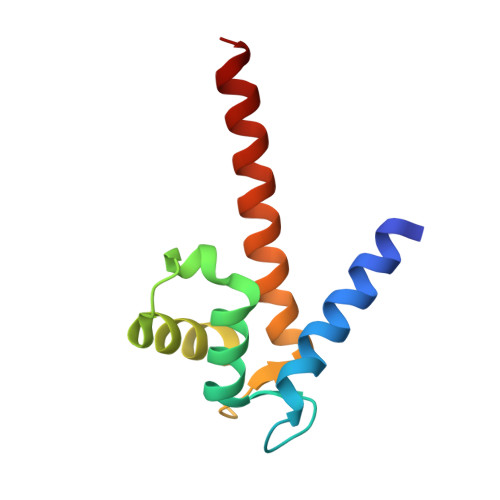
    2Y5I

  • PubMed Abstract: 

    The S100 family, with about 20 members in humans, is composed of EF-hand calcium-regulated proteins and is linked to a range of serious human diseases, including cancer and autoimmune and neurological disorders. The oldest S100 family members are found in teleosts (bony fish). The zebrafish, Danio rerio, was suggested as a promising model system for in vivo studies on S100 family functions, and we chose to investigate zebrafish S100Z as the closest homologue of the metastasis-promoting human S100A4. Here, we report the first crystal structure of an S100 protein from this organism, the calcium-bound state of S100Z to 2.03 Å resolution. Crystal packing suggests higher-order oligomerisation of S100Z dimers, with a tetramerisation interface very similar to, but even more extensive than, that reported for S100A4. The interactions are primarily through the C-terminal αIV helices from adjacent dimers in an antiparallel orientation. Structural comparisons between known S100 multimeric assemblies together with analysis of calcium-driven changes to the dimerisation cores suggest a mechanism for calcium-promoted oligomerisation of S100 proteins.


  • Organizational Affiliation
    • Structural Biology Laboratory, Department of Chemistry, University of York, Heslington, York YO10 5DD, UK. olga@ysbl.york.ac.uk

Macromolecule Content 

  • Total Structure Weight: 68.35 kDa 
  • Atom Count: 5,238 
  • Modeled Residue Count: 584 
  • Deposited Residue Count: 594 
  • Unique protein chains: 1

Macromolecules

Find similar proteins by:|  3D Structure
Entity ID: 1
MoleculeChains  Sequence LengthOrganismDetailsImage
S100 CALCIUM BINDING PROTEIN Z
A, B, C, D, E
A, B, C, D, E, F
99Danio rerioMutation(s): 0 
UniProt
Find proteins for Q503K9 (Danio rerio)
Explore Q503K9 
Go to UniProtKB:  Q503K9
Entity Groups
Sequence Clusters30% Identity50% Identity70% Identity90% Identity95% Identity100% Identity
UniProt GroupQ503K9
Sequence Annotations
Expand
Reference Sequence

Small Molecules

Ligands 2 Unique
IDChains Name / Formula / InChI Key2D Diagram3D Interactions
IPA

Query on IPA


Download:Ideal Coordinates CCD File
I [auth A]
N [auth C]
Q [auth D]
T [auth E]
W [auth F]
I [auth A],
N [auth C],
Q [auth D],
T [auth E],
W [auth F],
X [auth F]
ISOPROPYL ALCOHOL
C3 H8 O
KFZMGEQAYNKOFK-UHFFFAOYSA-N
CA

Query on CA


Download:Ideal Coordinates CCD File
G [auth A]
H [auth A]
J [auth B]
K [auth B]
L [auth C]
G [auth A],
H [auth A],
J [auth B],
K [auth B],
L [auth C],
M [auth C],
O [auth D],
P [auth D],
R [auth E],
S [auth E],
U [auth F],
V [auth F]
CALCIUM ION
Ca
BHPQYMZQTOCNFJ-UHFFFAOYSA-N

Experimental Data & Validation

Experimental Data

  • Method: X-RAY DIFFRACTION
  • Resolution: 2.03 Å
  • R-Value Free:  0.227 (Depositor), 0.206 (DCC) 
  • R-Value Work:  0.171 (Depositor), 0.179 (DCC) 
  • R-Value Observed: 0.174 (Depositor) 
Space Group: P 21 21 2
Unit Cell:
Length ( Å )Angle ( ˚ )
a = 75.611α = 90
b = 132.25β = 90
c = 58.209γ = 90
Software Package:
Software NamePurpose
REFMACrefinement
HKL-2000data reduction
SCALEPACKdata scaling
MOLREPphasing

Structure Validation

View Full Validation Report


Entry History 

Deposition Data

Revision History  (Full details and data files)

  • Version 1.0: 2011-07-27
    Type: Initial release
  • Version 1.1: 2011-08-24
    Changes: Database references
  • Version 1.2: 2023-12-20
    Changes: Data collection, Database references, Derived calculations, Other, Refinement description