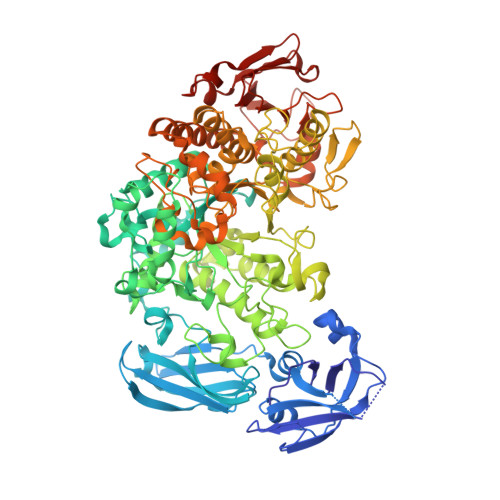
2Y5E

BARLEY LIMIT DEXTRINASE IN COMPLEX WITH ALPHA-CYCLODEXTRIN


Experimental Data Snapshot

  • Method: X-RAY DIFFRACTION
  • Resolution: 2.49 Å
  • R-Value Free: 0.226 
  • R-Value Work: 0.175 
  • R-Value Observed: 0.175 

Starting Model: experimental
View more details

wwPDB Validation   3D Report Full Report


This is version 2.1 of the entry. See complete history


Literature

Crystal Structure of an Essential Enzyme in Seed Starch Degradation: Barley Limit Dextrinase in Complex with Cyclodextrins.

Vester-Christensen, M.B.Abou Hachem, M.Svensson, B.Henriksen, A.

(2010) J Mol Biol 403: 739

  • DOI: https://doi.org/10.1016/j.jmb.2010.09.031
  • Primary Citation of Related Structures:  
    2Y4S, 2Y5E

  • PubMed Abstract: 

    Barley limit dextrinase [Hordeum vulgare limit dextrinase (HvLD)] catalyzes the hydrolysis of α-1,6 glucosidic linkages in limit dextrins. This activity plays a role in starch degradation during germination and presumably in starch biosynthesis during grain filling. The crystal structures of HvLD in complex with the competitive inhibitors α-cyclodextrin (CD) and β-CD are solved and refined to 2.5 Å and 2.1 Å, respectively, and are the first structures of a limit dextrinase. HvLD belongs to glycoside hydrolase 13 family and is composed of four domains: an immunoglobulin-like N-terminal eight-stranded β-sandwich domain, a six-stranded β-sandwich domain belonging to the carbohydrate binding module 48 family, a catalytic (β/α)(8)-like barrel domain that lacks α-helix 5, and a C-terminal eight-stranded β-sandwich domain of unknown function. The CDs are bound at the active site occupying carbohydrate binding subsites +1 and +2. A glycerol and three water molecules mimic a glucose residue at subsite -1, thereby identifying residues involved in catalysis. The bulky Met440, a unique residue at its position among α-1,6 acting enzymes, obstructs subsite -4. The steric hindrance observed is proposed to affect substrate specificity and to cause a low activity of HvLD towards amylopectin. An extended loop (Asp513-Asn520) between β5 and β6 of the catalytic domain also seems to influence substrate specificity and to give HvLD a higher affinity for α-CD than pullulanases. The crystal structures additionally provide new insight into cation sites and the concerted action of the battery of hydrolytic enzymes in starch degradation.


  • Organizational Affiliation

    Enzyme and Protein Chemistry, Department of Systems Biology, Søltofts Plads, Building 224, Technical University of Denmark, Kgs. Lyngby, Denmark.


Macromolecules
Find similar proteins by:  (by identity cutoff)  |  3D Structure
Entity ID: 1
MoleculeChains Sequence LengthOrganismDetailsImage
LIMIT DEXTRINASE884Hordeum vulgareMutation(s): 3 
EC: 3.2.1.41
UniProt
Find proteins for O48541 (Hordeum vulgare subsp. vulgare)
Explore O48541 
Go to UniProtKB:  O48541
Entity Groups  
Sequence Clusters30% Identity50% Identity70% Identity90% Identity95% Identity100% Identity
UniProt GroupO48541
Sequence Annotations
Expand
  • Reference Sequence
Oligosaccharides

Help

Entity ID: 2
MoleculeChains Length2D Diagram Glycosylation3D Interactions
Cyclohexakis-(1-4)-(alpha-D-glucopyranose)
B
6N/A
Glycosylation Resources
GlyTouCan:  G22307HL
GlyCosmos:  G22307HL
Small Molecules
Ligands 3 Unique
IDChains Name / Formula / InChI Key2D Diagram3D Interactions
IOD
Query on IOD

Download Ideal Coordinates CCD File 
F [auth A]
G [auth A]
H [auth A]
I [auth A]
J [auth A]
F [auth A],
G [auth A],
H [auth A],
I [auth A],
J [auth A],
K [auth A],
L [auth A],
M [auth A]
IODIDE ION
I
XMBWDFGMSWQBCA-UHFFFAOYSA-M
GOL
Query on GOL

Download Ideal Coordinates CCD File 
C [auth A]GLYCEROL
C3 H8 O3
PEDCQBHIVMGVHV-UHFFFAOYSA-N
CA
Query on CA

Download Ideal Coordinates CCD File 
D [auth A],
E [auth A]
CALCIUM ION
Ca
BHPQYMZQTOCNFJ-UHFFFAOYSA-N
Biologically Interesting Molecules (External Reference) 1 Unique
Experimental Data & Validation

Experimental Data

  • Method: X-RAY DIFFRACTION
  • Resolution: 2.49 Å
  • R-Value Free: 0.226 
  • R-Value Work: 0.175 
  • R-Value Observed: 0.175 
  • Space Group: C 1 2 1
Unit Cell:
Length ( Å )Angle ( ˚ )
a = 174.84α = 90
b = 85.61β = 96.83
c = 61.12γ = 90
Software Package:
Software NamePurpose
PHENIXrefinement
MOSFLMdata reduction
SCALAdata scaling
PHASERphasing

Structure Validation

View Full Validation Report


Entry History 

Deposition Data

Revision History  (Full details and data files)

  • Version 1.0: 2011-01-26
    Type: Initial release
  • Version 1.1: 2012-11-21
    Changes: Database references, Non-polymer description
  • Version 1.2: 2018-01-17
    Changes: Data collection
  • Version 2.0: 2020-07-29
    Type: Remediation
    Reason: Carbohydrate remediation
    Changes: Atomic model, Data collection, Derived calculations, Non-polymer description, Other, Structure summary
  • Version 2.1: 2023-12-20
    Changes: Data collection, Database references, Refinement description, Structure summary