2Y4N | pdb_00002y4n

PaaK1 in complex with phenylacetyl adenylate


Experimental Data Snapshot

  • Method: X-RAY DIFFRACTION
  • Resolution: 1.92 Å
  • R-Value Free: 
    0.232 (Depositor), 0.229 (DCC) 
  • R-Value Work: 
    0.181 (Depositor), 0.180 (DCC) 
  • R-Value Observed: 
    0.183 (Depositor) 

Starting Model: experimental
View more details

wwPDB Validation 3D Report Full Report

Validation slider image for 2Y4N

Ligand Structure Quality Assessment 


This is version 1.4 of the entry. See complete history

Literature

Defining a Structural and Kinetic Rationale for Paralogous Copies of Phenylacetate-Coa Ligases from the Cystic Fibrosis Pathogen Burkholderia Cenocepacia J2315.

Law, A.Boulanger, M.J.

(2011) J Biological Chem 286: 15577

  • DOI: https://doi.org/10.1074/jbc.M111.219683
  • Primary Citation Related Structures: 
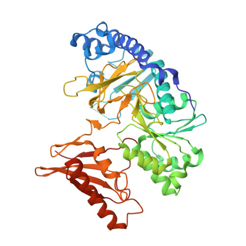
    2Y27, 2Y4N, 2Y4O

  • PubMed Abstract: 

    The phenylacetic acid (PAA) degradation pathway is the sole aerobic route for phenylacetic acid metabolism in bacteria and facilitates degradation of environmental pollutants such as styrene and ethylbenzene. The PAA pathway also is implicated in promoting Burkholderia cenocepacia infections in cystic fibrosis patients. Intriguingly, the first enzyme in the PAA pathway is present in two copies (paaK1 and paaK2), yet each subsequent enzyme is present in only a single copy. Furthermore, sequence divergence indicates that PaaK1 and PaaK2 form a unique subgroup within the adenylate-forming enzyme (AFE) superfamily. To establish a biochemical rationale for the existence of the PaaK paralogs in B. cenocepacia, we present high resolution x-ray crystal structures of a selenomethionine derivative of PaaK1 in complex with ATP and adenylated phenylacetate intermediate complexes of PaaK1 and PaaK2 in distinct conformations. Structural analysis reveals a novel N-terminal microdomain that may serve to recruit subsequent PAA enzymes, whereas a bifunctional role is proposed for the P-loop in stabilizing the C-terminal domain in conformation 2. The potential for different kinetic profiles was suggested by a structurally divergent extension of the aryl substrate pocket in PaaK1 relative to PaaK2. Functional characterization confirmed this prediction, with PaaK1 possessing a lower K(m) for phenylacetic acid and better able to accommodate 3' and 4' substitutions on the phenyl ring. Collectively, these results offer detailed insight into the reaction mechanism of a novel subgroup of the AFE superfamily and provide a clear biochemical rationale for the presence of paralogous copies of PaaK of B. cenocepacia.


  • Organizational Affiliation
    • Department of Biochemistry & Microbiology, University of Victoria, Victoria, British Columbia V8W 3P6, Canada.

Macromolecule Content 

  • Total Structure Weight: 98.04 kDa 
  • Atom Count: 7,400 
  • Modeled Residue Count: 849 
  • Deposited Residue Count: 874 
  • Unique protein chains: 1

Macromolecules

Find similar proteins by:|  3D Structure
Entity ID: 1
MoleculeChains  Sequence LengthOrganismDetailsImage
PHENYLACETATE-COENZYME A LIGASE
A, B
437Burkholderia cenocepacia J2315Mutation(s): 0 
EC: 6.2.1.30
UniProt
Find proteins for B4E7B5 (Burkholderia cenocepacia (strain ATCC BAA-245 / DSM 16553 / LMG 16656 / NCTC 13227 / J2315 / CF5610))
Explore B4E7B5 
Go to UniProtKB:  B4E7B5
Entity Groups
Sequence Clusters30% Identity50% Identity70% Identity90% Identity95% Identity100% Identity
UniProt GroupB4E7B5
Sequence Annotations
Expand
Reference Sequence

Small Molecules

Ligands 6 Unique
IDChains Name / Formula / InChI Key2D Diagram3D Interactions
DLL

Query on DLL


Download:Ideal Coordinates CCD File
G [auth A],
N [auth B]
5'-O-[HYDROXY(PHENYLACETYL)PHOSPHORYL]ADENOSINE
C18 H20 N5 O8 P
VUKDZGAUWUDQRZ-XKLVTHTNSA-N
PGE

Query on PGE


Download:Ideal Coordinates CCD File
K [auth B]TRIETHYLENE GLYCOL
C6 H14 O4
ZIBGPFATKBEMQZ-UHFFFAOYSA-N
PAC

Query on PAC


Download:Ideal Coordinates CCD File
J [auth B]2-PHENYLACETIC ACID
C8 H8 O2
WLJVXDMOQOGPHL-UHFFFAOYSA-N
PEG

Query on PEG


Download:Ideal Coordinates CCD File
E [auth A]DI(HYDROXYETHYL)ETHER
C4 H10 O3
MTHSVFCYNBDYFN-UHFFFAOYSA-N
BME

Query on BME


Download:Ideal Coordinates CCD File
F [auth A],
L [auth B],
M [auth B]
BETA-MERCAPTOETHANOL
C2 H6 O S
DGVVWUTYPXICAM-UHFFFAOYSA-N
MG

Query on MG


Download:Ideal Coordinates CCD File
C [auth A],
D [auth A],
H [auth B],
I [auth B]
MAGNESIUM ION
Mg
JLVVSXFLKOJNIY-UHFFFAOYSA-N

Experimental Data & Validation

Experimental Data

  • Method: X-RAY DIFFRACTION
  • Resolution: 1.92 Å
  • R-Value Free:  0.232 (Depositor), 0.229 (DCC) 
  • R-Value Work:  0.181 (Depositor), 0.180 (DCC) 
  • R-Value Observed: 0.183 (Depositor) 
Space Group: P 1
Unit Cell:
Length ( Å )Angle ( ˚ )
a = 57.26α = 91.16
b = 62.39β = 108.33
c = 76.59γ = 106.27
Software Package:
Software NamePurpose
REFMACrefinement
iMOSFLMdata reduction
SCALAdata scaling
MOLREPphasing

Structure Validation

View Full Validation Report


Ligand Structure Quality Assessment 


Entry History 

Deposition Data

Revision History  (Full details and data files)

  • Version 1.0: 2011-03-01
    Type: Initial release
  • Version 1.1: 2011-05-08
    Changes: Version format compliance
  • Version 1.2: 2011-07-13
    Changes: Version format compliance
  • Version 1.3: 2017-07-12
    Changes: Derived calculations
  • Version 1.4: 2023-12-20
    Changes: Data collection, Database references, Derived calculations, Other, Refinement description