How the Biotin-Streptavidin Interaction Was Made Even Stronger: Investigation Via Crystallography and a Chimeric Tetramer.
Chivers, C.E., Koner, A.L., Lowe, E.D., Howarth, M.(2011) Biochem J 435: 55
- PubMed: 21241253 Search on PubMedSearch on PubMed Central
- DOI: https://doi.org/10.1042/BJ20101593
- Primary Citation Related Structures:
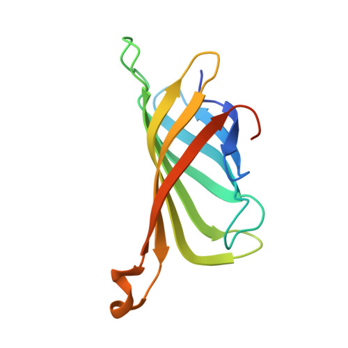
2Y3E, 2Y3F - PubMed Abstract:
The interaction between SA (streptavidin) and biotin is one of the strongest non-covalent interactions in Nature. SA is a widely used tool and a paradigm for protein-ligand interactions. We previously developed a SA mutant, termed Tr (traptavidin), possessing a 10-fold lower off-rate for biotin, with increased mechanical and thermal stability. In the present study, we determined the crystal structures of apo-Tr and biotin-Tr at 1.5 Å resolution. In apo-SA the loop (L3/4), near biotin's valeryl tail, is typically disordered and open, but closes upon biotin binding. In contrast, L3/4 was shut in both apo-Tr and biotin-Tr. The reduced flexibility of L3/4 and decreased conformational change on biotin binding provide an explanation for Tr's reduced biotin off- and on-rates. L3/4 includes Ser45, which forms a hydrogen bond to biotin consistently in Tr, but erratically in SA. Reduced breakage of the biotin-Ser45 hydrogen bond in Tr is likely to inhibit the initiating event in biotin's dissociation pathway. We generated a Tr with a single biotin-binding site rather than four, which showed a simi-larly low off-rate, demonstrating that Tr's low off-rate was governed by intrasubunit effects. Understanding the structural features of this tenacious interaction may assist the design of even stronger affinity tags and inhibitors.
- Department of Biochemistry, Oxford University, South Parks Road, Oxford OX1 3QU, UK.
Organizational Affiliation: