2Y24

STRUCTURAL BASIS FOR SUBSTRATE RECOGNITION BY ERWINIA CHRYSANTHEMI GH5 GLUCURONOXYLANASE


Experimental Data Snapshot

  • Method: X-RAY DIFFRACTION
  • Resolution: 1.39 Å
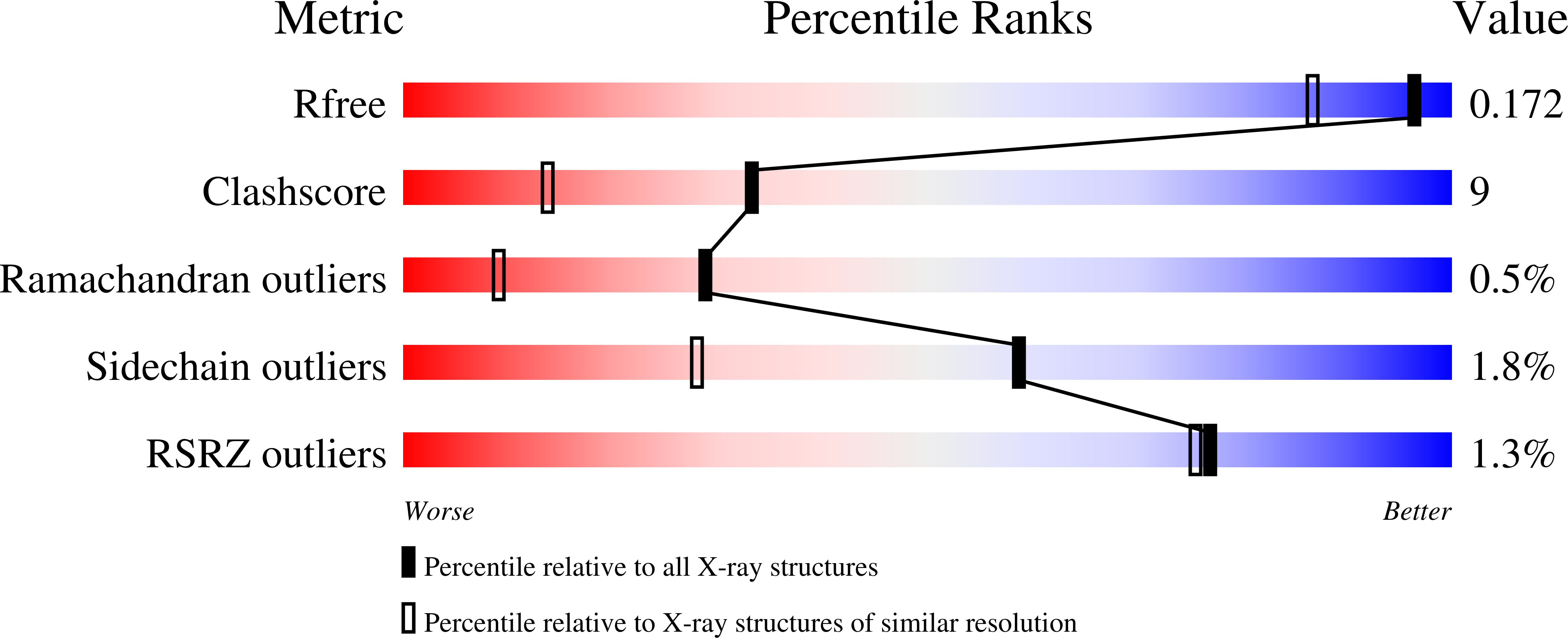
  • R-Value Free: 0.169 
  • R-Value Work: 0.120 
  • R-Value Observed: 0.122 

Starting Model: experimental
View more details

wwPDB Validation   3D Report Full Report


This is version 2.1 of the entry. See complete history


Literature

Structural Basis for Substrate Recognition by Erwinia Chrysanthemi Gh30 Glucuronoxylanase.

Urbanikova, L.Vrsanska, M.Krogh, K.B.Hoff, T.Biely, P.

(2011) FEBS J 278: 2105

  • DOI: https://doi.org/10.1111/j.1742-4658.2011.08127.x
  • Primary Citation of Related Structures:  
    2Y24

  • PubMed Abstract: 

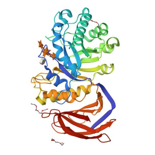
    Xylanase A from the phytopathogenic bacterium Erwinia chrysanthemi is classified as a glycoside hydrolase family 30 enzyme (previously in family 5) and is specialized for degradation of glucuronoxylan. The recombinant enzyme was crystallized with the aldotetraouronic acid β-D-xylopyranosyl-(1→4)-[4-O-methyl-α-D-glucuronosyl-(1→2)]-β-D-xylopyranosyl-(1→4)-D-xylose as a ligand. The crystal structure of the enzyme-ligand complex was solved at 1.39 Å resolution. The ligand xylotriose moiety occupies subsites -1, -2 and -3, whereas the methyl glucuronic acid residue attached to the middle xylopyranosyl residue of xylotriose is bound to the enzyme through hydrogen bonds to five amino acids and by the ionic interaction of the methyl glucuronic acid carboxylate with the positively charged guanidinium group of Arg293. The interaction of the enzyme with the methyl glucuronic acid residue appears to be indispensable for proper distortion of the xylan chain and its effective hydrolysis. Such a distortion does not occur with linear β-1,4-xylooligosaccharides, which are hydrolyzed by the enzyme at a negligible rate. Structural and experimental data are available in the Protein Data Bank database under accession number 2y24 [45].


  • Organizational Affiliation

    Institute of Molecular Biology, Slovak Academy of Sciences, Bratislava, Slovakia.


Macromolecules
Find similar proteins by:  (by identity cutoff)  |  3D Structure
Entity ID: 1
MoleculeChains Sequence LengthOrganismDetailsImage
XYLANASE383Dickeya chrysanthemiMutation(s): 0 
EC: 3.2.1.8 (PDB Primary Data), 3.2.1.136 (PDB Primary Data)
UniProt
Find proteins for Q46961 (Dickeya chrysanthemi)
Explore Q46961 
Go to UniProtKB:  Q46961
Entity Groups  
Sequence Clusters30% Identity50% Identity70% Identity90% Identity95% Identity100% Identity
UniProt GroupQ46961
Sequence Annotations
Expand
  • Reference Sequence
Oligosaccharides

Help

Entity ID: 2
MoleculeChains Length2D Diagram Glycosylation3D Interactions
4-O-methyl-alpha-D-glucopyranuronic acid-(1-2)-[beta-D-xylopyranose-(1-4)]beta-D-xylopyranose-(1-4)-beta-D-xylopyranose
B
4N/A
Glycosylation Resources
GlyTouCan:  G14569KK
GlyCosmos:  G14569KK
Experimental Data & Validation

Experimental Data

  • Method: X-RAY DIFFRACTION
  • Resolution: 1.39 Å
  • R-Value Free: 0.169 
  • R-Value Work: 0.120 
  • R-Value Observed: 0.122 
  • Space Group: P 32 2 1
Unit Cell:
Length ( Å )Angle ( ˚ )
a = 59.578α = 90
b = 59.578β = 90
c = 168.296γ = 120
Software Package:
Software NamePurpose
REFMACrefinement
XDSdata reduction
SCALEPACKdata scaling
MOLREPphasing

Structure Validation

View Full Validation Report


Entry History 

Deposition Data

Revision History  (Full details and data files)

  • Version 1.0: 2011-06-01
    Type: Initial release
  • Version 1.1: 2011-12-21
    Changes: Database references, Version format compliance
  • Version 1.2: 2019-07-17
    Changes: Data collection, Derived calculations
  • Version 2.0: 2020-07-29
    Type: Remediation
    Reason: Carbohydrate remediation
    Changes: Atomic model, Data collection, Derived calculations, Other, Structure summary
  • Version 2.1: 2023-12-20
    Changes: Data collection, Database references, Derived calculations, Refinement description, Structure summary