2Y1A | pdb_00002y1a

Crystal structure of Achromobacter cycloclastes Cu nitrite reductase with bound NO


Experimental Data Snapshot

  • Method: X-RAY DIFFRACTION
  • Resolution: 1.95 Å
  • R-Value Free: 
    0.210 (Depositor), 0.208 (DCC) 
  • R-Value Work: 
    0.153 (Depositor), 0.162 (DCC) 
  • R-Value Observed: 
    0.156 (Depositor) 

Starting Model: experimental
View more details

wwPDB Validation 3D Report Full Report

Validation slider image for 2Y1A

This is version 1.3 of the entry. See complete history

Literature

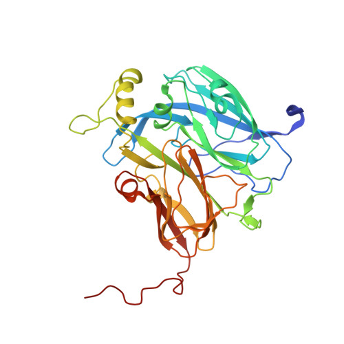
Monitoring and Validating Active Site Redox States in Protein Crystals.

Antonyuk, S.V.Hough, M.A.

(2011) Biochim Biophys Acta 1814: 778

  • DOI: https://doi.org/10.1016/j.bbapap.2010.12.017
  • Primary Citation Related Structures: 
    2Y1A, 2Y3Q

  • PubMed Abstract: 

    High resolution protein crystallography using synchrotron radiation is one of the most powerful tools in modern biology. Improvements in resolution have arisen from the use of X-ray beamlines with higher brightness and flux and the development of advanced detectors. However, it is increasingly recognised that the benefits brought by these advances have an associated cost, namely deleterious effects of X-ray radiation on the sample (radiation damage). In particular, X-ray induced reduction and damage to redox centres has been shown to occur much more rapidly than other radiation damage effects, such as loss of resolution or damage to disulphide bridges. Selection of an appropriate combination of in-situ single crystal spectroscopies during crystallographic experiments, such as UV-visible absorption and X-ray absorption spectroscopy (XAFS), allows for effective monitoring of redox states in protein crystals in parallel with structure determination. Such approaches are also essential in cases where catalytic intermediate species are generated by exposure to the X-ray beam. In this article, we provide a number of examples in which multiple single crystal spectroscopies have been key to understanding the redox status of Fe and Cu centres in crystal structures. This article is part of a Special Issue entitled: Protein Structure and Function in the Crystalline State.


  • Organizational Affiliation
    • Faculty of Health and Life Sciences, University of Liverpool, Crown Street, Liverpool L69 7ZB, UK.

Macromolecule Content 

  • Total Structure Weight: 37.72 kDa 
  • Atom Count: 2,845 
  • Modeled Residue Count: 332 
  • Deposited Residue Count: 340 
  • Unique protein chains: 1

Macromolecules

Find similar proteins by:|  3D Structure
Entity ID: 1
MoleculeChains  Sequence LengthOrganismDetailsImage
COPPER-CONTAINING NITRITE REDUCTASE340Achromobacter cycloclastesMutation(s): 0 
EC: 1.7.2.1
UniProt
Find proteins for P25006 (Achromobacter cycloclastes)
Explore P25006 
Go to UniProtKB:  P25006
Entity Groups
Sequence Clusters30% Identity50% Identity70% Identity90% Identity95% Identity100% Identity
UniProt GroupP25006
Sequence Annotations
Expand
Reference Sequence

Small Molecules

Ligands 4 Unique
IDChains Name / Formula / InChI Key2D Diagram3D Interactions
SO4

Query on SO4


Download:Ideal Coordinates CCD File
G [auth A],
H [auth A],
I [auth A],
J [auth A]
SULFATE ION
O4 S
QAOWNCQODCNURD-UHFFFAOYSA-L
CU

Query on CU


Download:Ideal Coordinates CCD File
B [auth A],
C [auth A]
COPPER (II) ION
Cu
JPVYNHNXODAKFH-UHFFFAOYSA-N
ACT

Query on ACT


Download:Ideal Coordinates CCD File
E [auth A],
F [auth A]
ACETATE ION
C2 H3 O2
QTBSBXVTEAMEQO-UHFFFAOYSA-M
NO

Query on NO


Download:Ideal Coordinates CCD File
D [auth A]NITRIC OXIDE
N O
ODUCDPQEXGNKDN-UHFFFAOYSA-N

Experimental Data & Validation

Experimental Data

  • Method: X-RAY DIFFRACTION
  • Resolution: 1.95 Å
  • R-Value Free:  0.210 (Depositor), 0.208 (DCC) 
  • R-Value Work:  0.153 (Depositor), 0.162 (DCC) 
  • R-Value Observed: 0.156 (Depositor) 
Space Group: P 21 3
Unit Cell:
Length ( Å )Angle ( ˚ )
a = 95.24α = 90
b = 95.24β = 90
c = 95.24γ = 90
Software Package:
Software NamePurpose
REFMACrefinement
HKL-2000data reduction
HKL-2000data scaling
MOLREPphasing

Structure Validation

View Full Validation Report


Entry History 

Deposition Data

Revision History  (Full details and data files)

  • Version 1.0: 2011-02-16
    Type: Initial release
  • Version 1.1: 2011-05-19
    Changes: Version format compliance
  • Version 1.2: 2011-07-13
    Changes: Version format compliance
  • Version 1.3: 2023-12-20
    Changes: Data collection, Database references, Derived calculations, Other, Refinement description