2XYD | pdb_00002xyd

human Angiotenisn converting enzyme N-domain in complex with Phosphinic tripeptide


Experimental Data Snapshot

  • Method: X-RAY DIFFRACTION
  • Resolution: 2.15 Å
  • R-Value Free: 
    0.256 (Depositor), 0.253 (DCC) 
  • R-Value Work: 
    0.220 (Depositor), 0.220 (DCC) 
  • R-Value Observed: 
    0.222 (Depositor) 

Starting Model: experimental
View more details

wwPDB Validation 3D Report Full Report

Validation slider image for 2XYD

Ligand Structure Quality Assessment 


This is version 2.2 of the entry. See complete history

Literature

Novel Mechanism of Inhibition of Human Angiotensin-I-Converting Enzyme (Ace) by a Highly Specific Phosphinic Tripeptide.

Akif, M.Schwager, S.L.Anthony, C.S.Czarny, B.Beau, F.Dive, V.Sturrock, E.D.Acharya, K.R.

(2011) Biochem J 436: 53

  • DOI: https://doi.org/10.1042/BJ20102123
  • Primary Citation Related Structures: 
    2XY9, 2XYD

  • PubMed Abstract: 

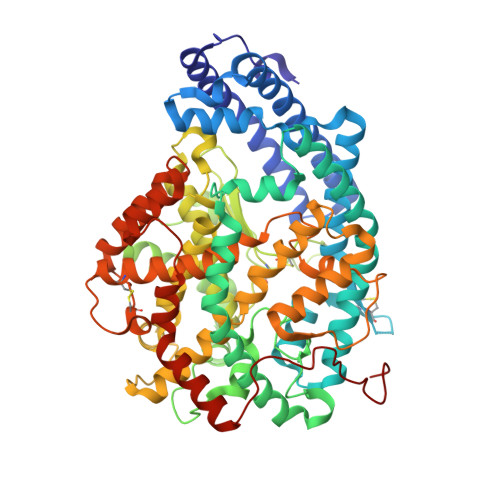
    Human ACE (angiotensin-I-converting enzyme) has long been regarded as an excellent target for the treatment of hypertension and related cardiovascular diseases. Highly potent inhibitors have been developed and are extensively used in the clinic. To develop inhibitors with higher therapeutic efficacy and reduced side effects, recent efforts have been directed towards the discovery of compounds able to simultaneously block more than one zinc metallopeptidase (apart from ACE) involved in blood pressure regulation in humans, such as neprilysin and ECE-1 (endothelin-converting enzyme-1). In the present paper, we show the first structures of testis ACE [C-ACE, which is identical with the C-domain of somatic ACE and the dominant domain responsible for blood pressure regulation, at 1.97Å (1 Å=0.1 nm)] and the N-domain of somatic ACE (N-ACE, at 2.15Å) in complex with a highly potent and selective dual ACE/ECE-1 inhibitor. The structural determinants revealed unique features of the binding of two molecules of the dual inhibitor in the active site of C-ACE. In both structures, the first molecule is positioned in the obligatory binding site and has a bulky bicyclic P(1)' residue with the unusual R configuration which, surprisingly, is accommodated by the large S(2)' pocket. In the C-ACE complex, the isoxazole phenyl group of the second molecule makes strong pi-pi stacking interactions with the amino benzoyl group of the first molecule locking them in a 'hand-shake' conformation. These features, for the first time, highlight the unusual architecture and flexibility of the active site of C-ACE, which could be further utilized for structure-based design of new C-ACE or vasopeptidase inhibitors.


  • Organizational Affiliation
    • Department of Biology and Biochemistry, University of Bath, Claverton Down, Bath BA2 7AY, UK.

Macromolecule Content 

  • Total Structure Weight: 146.47 kDa 
  • Atom Count: 10,490 
  • Modeled Residue Count: 1,214 
  • Deposited Residue Count: 1,220 
  • Unique protein chains: 1

Macromolecules

Find similar proteins by:|  3D Structure
Entity ID: 1
MoleculeChains  Sequence LengthOrganismDetailsImage
ANGIOTENSIN-CONVERTING ENZYME
A, B
610Homo sapiensMutation(s): 7 
EC: 3.4.15.1
UniProt & NIH Common Fund Data Resources
Find proteins for P12821 (Homo sapiens)
Explore P12821 
Go to UniProtKB:  P12821
PHAROS:  P12821
GTEx:  ENSG00000159640 
Entity Groups
Sequence Clusters30% Identity50% Identity70% Identity90% Identity95% Identity100% Identity
UniProt GroupP12821
Glycosylation
Glycosylation Sites: 3Go to GlyGen: P12821-1
Sequence Annotations
Expand
Reference Sequence

Oligosaccharides

Help  
Entity ID: 2
MoleculeChains Length2D Diagram GlycosylationD Interactions
alpha-L-fucopyranose-(1-6)-2-acetamido-2-deoxy-beta-D-glucopyranose
C, F
2N-Glycosylation
Glycosylation Resources
GlyTouCan: G86851RC
GlyCosmos: G86851RC
GlyGen: G86851RC
Entity ID: 3
MoleculeChains Length2D Diagram GlycosylationD Interactions
2-acetamido-2-deoxy-beta-D-glucopyranose-(1-4)-2-acetamido-2-deoxy-beta-D-glucopyranose
D, G
2N-Glycosylation
Glycosylation Resources
GlyTouCan: G42666HT
GlyCosmos: G42666HT
GlyGen: G42666HT
Entity ID: 4
MoleculeChains Length2D Diagram GlycosylationD Interactions
beta-D-mannopyranose-(1-4)-2-acetamido-2-deoxy-beta-D-glucopyranose-(1-4)-[alpha-L-fucopyranose-(1-6)]2-acetamido-2-deoxy-beta-D-glucopyranose
E
4N-Glycosylation
Glycosylation Resources
GlyTouCan: G32152BH
GlyCosmos: G32152BH
GlyGen: G32152BH
Entity ID: 5
MoleculeChains Length2D Diagram GlycosylationD Interactions
beta-D-mannopyranose-(1-4)-2-acetamido-2-deoxy-beta-D-glucopyranose-(1-4)-2-acetamido-2-deoxy-beta-D-glucopyranose
H
3N-Glycosylation
Glycosylation Resources
GlyTouCan: G15407YE
GlyCosmos: G15407YE
GlyGen: G15407YE

Small Molecules

Ligands 6 Unique
IDChains Name / Formula / InChI Key2D Diagram3D Interactions
3ES

Query on 3ES


Download:Ideal Coordinates CCD File
I [auth A],
P [auth B]
[(2S)-2-({3-[HYDROXYL(2-PHENYL-(1R)-1-{[(BENZYLOXY)[(2S)-2-({3-[HYDROXYL(2-PHENYL-(1R)-1-CARBONYL]-AMINO}ETHYL)PHOSPHINYL]-2-[(3-PHENYLISOXAZOL-5-YL)METHYL]-1-OXO-PROPYL}AMINO)-3-(4-HYDROXY-PHENYL)
C38 H38 N3 O9 P
ZPFSKFCSVXPMBD-PFESQZPFSA-N
P6G

Query on P6G


Download:Ideal Coordinates CCD File
T [auth B]HEXAETHYLENE GLYCOL
C12 H26 O7
IIRDTKBZINWQAW-UHFFFAOYSA-N
PG4

Query on PG4


Download:Ideal Coordinates CCD File
O [auth A],
S [auth B]
TETRAETHYLENE GLYCOL
C8 H18 O5
UWHCKJMYHZGTIT-UHFFFAOYSA-N
PEG

Query on PEG


Download:Ideal Coordinates CCD File
L [auth A],
M [auth A],
N [auth A],
U [auth B]
DI(HYDROXYETHYL)ETHER
C4 H10 O3
MTHSVFCYNBDYFN-UHFFFAOYSA-N
ZN

Query on ZN


Download:Ideal Coordinates CCD File
J [auth A],
Q [auth B]
ZINC ION
Zn
PTFCDOFLOPIGGS-UHFFFAOYSA-N
CL

Query on CL


Download:Ideal Coordinates CCD File
K [auth A],
R [auth B]
CHLORIDE ION
Cl
VEXZGXHMUGYJMC-UHFFFAOYSA-M

Experimental Data & Validation

Experimental Data

  • Method: X-RAY DIFFRACTION
  • Resolution: 2.15 Å
  • R-Value Free:  0.256 (Depositor), 0.253 (DCC) 
  • R-Value Work:  0.220 (Depositor), 0.220 (DCC) 
  • R-Value Observed: 0.222 (Depositor) 
Space Group: P 1
Unit Cell:
Length ( Å )Angle ( ˚ )
a = 72.837α = 89.11
b = 76.437β = 64.43
c = 82.954γ = 75.87
Software Package:
Software NamePurpose
REFMACrefinement
HKL-2000data reduction
SCALEPACKdata scaling
PHASERphasing

Structure Validation

View Full Validation Report


Ligand Structure Quality Assessment 


Entry History 

Deposition Data

Revision History  (Full details and data files)

  • Version 1.0: 2011-05-11
    Type: Initial release
  • Version 1.1: 2012-04-11
    Changes: Non-polymer description, Version format compliance
  • Version 2.0: 2020-07-29
    Type: Remediation
    Reason: Carbohydrate remediation
    Changes: Advisory, Atomic model, Data collection, Derived calculations, Other, Structure summary
  • Version 2.1: 2023-12-20
    Changes: Data collection, Database references, Refinement description, Structure summary
  • Version 2.2: 2024-10-23
    Changes: Structure summary