2XYA | pdb_00002xya

Non-covalent inhibtors of rhinovirus 3C protease.


Experimental Data Snapshot

  • Method: X-RAY DIFFRACTION
  • Resolution: 2.40 Å
  • R-Value Free: 
    0.288 (Depositor) 
  • R-Value Work: 
    0.227 (Depositor), 0.217 (DCC) 
  • R-Value Observed: 
    0.230 (Depositor) 

wwPDB Validation 3D Report Full Report

Validation slider image for 2XYA

Ligand Structure Quality Assessment 


This is version 1.3 of the entry. See complete history

Literature

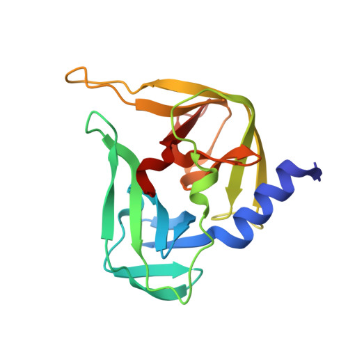
Non-Covalent Inhibitors of Rhinovirus 3C Protease.

Baxter, A.Chambers, M.Edfeldt, F.Edman, K.Freeman, A.Johansson, C.King, S.Morley, A.Petersen, J.Rawlins, P.Spadola, L.Thong, B.Poel, H.V.Williams, N.

(2011) Bioorg Med Chem Lett 21: 777

  • DOI: https://doi.org/10.1016/j.bmcl.2010.11.110
  • Primary Citation Related Structures: 
    2XYA

  • PubMed Abstract: 

    The first known non-covalent inhibitors of rhinovirus 3C protease (3CP) have been identified through fragment based screening and hit identification activities.


  • Organizational Affiliation
    • AstraZeneca, R&D Charnwood, Bakewell Road, Loughborough, Leicestershire LE11 5RH, UK.

Macromolecule Content 

  • Total Structure Weight: 20.35 kDa 
  • Atom Count: 1,462 
  • Modeled Residue Count: 182 
  • Deposited Residue Count: 182 
  • Unique protein chains: 1

Macromolecules

Find similar proteins by:|  3D Structure
Entity ID: 1
MoleculeChains  Sequence LengthOrganismDetailsImage
PICORNAIN 3C182Human rhinovirus sp.Mutation(s): 0 
EC: 3.4.22.28
UniProt
Find proteins for P04936 (Human rhinovirus 2)
Explore P04936 
Go to UniProtKB:  P04936
Entity Groups
Sequence Clusters30% Identity50% Identity70% Identity90% Identity95% Identity100% Identity
UniProt GroupP04936
Sequence Annotations
Expand
Reference Sequence

Small Molecules

Ligands 1 Unique
IDChains Name / Formula / InChI Key2D Diagram3D Interactions
7L4

Query on 7L4


Download:Ideal Coordinates CCD File
B [auth A]2-PHENYLQUINOLIN-4-OL
C15 H11 N O
JGABMVVOXLQCKZ-UHFFFAOYSA-N

Experimental Data & Validation

Experimental Data

  • Method: X-RAY DIFFRACTION
  • Resolution: 2.40 Å
  • R-Value Free:  0.288 (Depositor) 
  • R-Value Work:  0.227 (Depositor), 0.217 (DCC) 
  • R-Value Observed: 0.230 (Depositor) 
Space Group: I 41 2 2
Unit Cell:
Length ( Å )Angle ( ˚ )
a = 126.143α = 90
b = 126.143β = 90
c = 75.454γ = 90
Software Package:
Software NamePurpose
REFMACrefinement
MOSFLMdata reduction
SCALAdata scaling

Structure Validation

View Full Validation Report


Ligand Structure Quality Assessment 


Entry History 

Deposition Data

Revision History  (Full details and data files)

  • Version 1.0: 2011-04-27
    Type: Initial release
  • Version 1.1: 2011-05-08
    Changes: Version format compliance
  • Version 1.2: 2011-07-13
    Changes: Version format compliance
  • Version 1.3: 2024-05-08
    Changes: Data collection, Database references, Derived calculations, Other