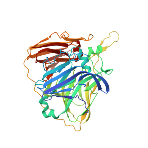
2XVB

Crystal structure of Laccase from Thermus thermophilus HB27 complexed with Hg, crystal of the apoenzyme soaked for 5 min. in 5 mM HgCl2 at 278 K.


Experimental Data Snapshot

  • Method: X-RAY DIFFRACTION
  • Resolution: 1.70 Å
  • R-Value Free: 0.178 
  • R-Value Work: 0.153 
  • R-Value Observed: 0.154 

Starting Model: experimental
View more details

wwPDB Validation   3D Report Full Report


This is version 1.3 of the entry. See complete history


Literature

X-Ray-Induced Catalytic Active-Site Reduction of a Multicopper Oxidase: Structural Insights Into the Proton-Relay Mechanism and O2-Reduction States.

Serrano-Posada, H.Centeno-Leija, S.Rojas-Trejo, S.P.Rodriguez-Almazan, C.Stojanoff, V.Rudino-Pinera, E.

(2015) Acta Crystallogr D Biol Crystallogr 71: 2396

  • DOI: https://doi.org/10.1107/S1399004715018714
  • Primary Citation of Related Structures:  
    2XU9, 2XUW, 2XVB, 2YAE, 2YAF, 2YAH, 2YAM, 2YAO, 2YAP, 2YAQ, 2YAR, 4AI7

  • PubMed Abstract: 

    During X-ray data collection from a multicopper oxidase (MCO) crystal, electrons and protons are mainly released into the system by the radiolysis of water molecules, leading to the X-ray-induced reduction of O2 to 2H2O at the trinuclear copper cluster (TNC) of the enzyme. In this work, 12 crystallographic structures of Thermus thermophilus HB27 multicopper oxidase (Tth-MCO) in holo, apo and Hg-bound forms and with different X-ray absorbed doses have been determined. In holo Tth-MCO structures with four Cu atoms, the proton-donor residue Glu451 involved in O2 reduction was found in a double conformation: Glu451a (∼7 Å from the TNC) and Glu451b (∼4.5 Å from the TNC). A positive peak of electron density above 3.5σ in an Fo - Fc map for Glu451a O(ℇ2) indicates the presence of a carboxyl functional group at the side chain, while its significant absence in Glu451b strongly suggests a carboxylate functional group. In contrast, for apo Tth-MCO and in Hg-bound structures neither the positive peak nor double conformations were observed. Together, these observations provide the first structural evidence for a proton-relay mechanism in the MCO family and also support previous studies indicating that Asp106 does not provide protons for this mechanism. In addition, eight composite structures (Tth-MCO-C1-8) with different X-ray-absorbed doses allowed the observation of different O2-reduction states, and a total depletion of T2Cu at doses higher than 0.2 MGy showed the high susceptibility of this Cu atom to radiation damage, highlighting the importance of taking radiation effects into account in biochemical interpretations of an MCO structure.


  • Organizational Affiliation

    Medicina Molecular y Bioprocesos, Instituto de Biotecnología, Universidad Nacional Autónoma de México, Avenida Universidad 2001, 62210 Cuernavaca, MOR, Mexico.


Macromolecules
Find similar proteins by:  (by identity cutoff)  |  3D Structure
Entity ID: 1
MoleculeChains Sequence LengthOrganismDetailsImage
LACCASE439Thermus thermophilus HB27Mutation(s): 0 
EC: 1.10.3.2
UniProt
Find proteins for Q72HW2 (Thermus thermophilus (strain ATCC BAA-163 / DSM 7039 / HB27))
Explore Q72HW2 
Go to UniProtKB:  Q72HW2
Entity Groups  
Sequence Clusters30% Identity50% Identity70% Identity90% Identity95% Identity100% Identity
UniProt GroupQ72HW2
Sequence Annotations
Expand
  • Reference Sequence
Small Molecules
Ligands 3 Unique
IDChains Name / Formula / InChI Key2D Diagram3D Interactions
HG
Query on HG

Download Ideal Coordinates CCD File 
B [auth A],
C [auth A],
D [auth A],
E [auth A],
F [auth A]
MERCURY (II) ION
Hg
BQPIGGFYSBELGY-UHFFFAOYSA-N
MRD
Query on MRD

Download Ideal Coordinates CCD File 
I [auth A]
J [auth A]
O [auth A]
Q [auth A]
V [auth A]
I [auth A],
J [auth A],
O [auth A],
Q [auth A],
V [auth A],
W [auth A]
(4R)-2-METHYLPENTANE-2,4-DIOL
C6 H14 O2
SVTBMSDMJJWYQN-RXMQYKEDSA-N
MPD
Query on MPD

Download Ideal Coordinates CCD File 
G [auth A]
H [auth A]
K [auth A]
L [auth A]
M [auth A]
G [auth A],
H [auth A],
K [auth A],
L [auth A],
M [auth A],
N [auth A],
P [auth A],
R [auth A],
S [auth A],
T [auth A],
U [auth A]
(4S)-2-METHYL-2,4-PENTANEDIOL
C6 H14 O2
SVTBMSDMJJWYQN-YFKPBYRVSA-N
Experimental Data & Validation

Experimental Data

  • Method: X-RAY DIFFRACTION
  • Resolution: 1.70 Å
  • R-Value Free: 0.178 
  • R-Value Work: 0.153 
  • R-Value Observed: 0.154 
  • Space Group: C 2 2 21
Unit Cell:
Length ( Å )Angle ( ˚ )
a = 93.456α = 90
b = 110.183β = 90
c = 96.343γ = 90
Software Package:
Software NamePurpose
PHENIXrefinement
XDSdata reduction
SCALAdata scaling
PHASERphasing

Structure Validation

View Full Validation Report


Entry History 

Deposition Data

Revision History  (Full details and data files)

  • Version 1.0: 2011-10-26
    Type: Initial release
  • Version 1.1: 2015-12-09
    Changes: Database references
  • Version 1.2: 2015-12-16
    Changes: Database references
  • Version 1.3: 2023-12-20
    Changes: Data collection, Database references, Derived calculations, Other, Refinement description