2XSE | pdb_00002xse

The structural basis for recognition of J-base containing DNA by a novel DNA-binding domain in JBP1


Experimental Data Snapshot

  • Method: X-RAY DIFFRACTION
  • Resolution: 1.90 Å
  • R-Value Free: 
    0.200 (Depositor), 0.214 (DCC) 
  • R-Value Work: 
    0.174 (Depositor), 0.189 (DCC) 
  • R-Value Observed: 
    0.176 (Depositor) 

wwPDB Validation 3D Report Full Report

Validation slider image for 2XSE

This is version 1.2 of the entry. See complete history

Literature

The Structural Basis for Recognition of Base J Containing DNA by a Novel DNA Binding Domain in Jbp1.

Heidebrecht, T.Christodoulou, E.Chalmers, M.J.Jan, S.Ter Riet, B.Grover, R.K.Joosten, R.P.Littler, D.Van Luenen, H.Griffin, P.R.Wentworth, P.Borst, P.Perrakis, A.

(2011) Nucleic Acids Res 39: 5715

  • DOI: https://doi.org/10.1093/nar/gkr125
  • Primary Citation Related Structures: 
    2XSE

  • PubMed Abstract: 

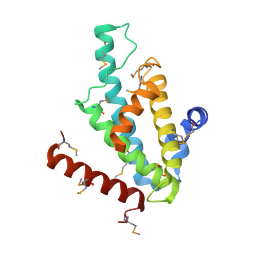
    The J-binding protein 1 (JBP1) is essential for biosynthesis and maintenance of DNA base-J (β-d-glucosyl-hydroxymethyluracil). Base-J and JBP1 are confined to some pathogenic protozoa and are absent from higher eukaryotes, prokaryotes and viruses. We show that JBP1 recognizes J-containing DNA (J-DNA) through a 160-residue domain, DB-JBP1, with 10 000-fold preference over normal DNA. The crystal structure of DB-JBP1 revealed a helix-turn-helix variant fold, a 'helical bouquet' with a 'ribbon' helix encompassing the amino acids responsible for DNA binding. Mutation of a single residue (Asp525) in the ribbon helix abrogates specificity toward J-DNA. The same mutation renders JBP1 unable to rescue the targeted deletion of endogenous JBP1 genes in Leishmania and changes its distribution in the nucleus. Based on mutational analysis and hydrogen/deuterium-exchange mass-spectrometry data, a model of JBP1 bound to J-DNA was constructed and validated by small-angle X-ray scattering data. Our results open new possibilities for targeted prevention of J-DNA recognition as a therapeutic intervention for parasitic diseases.


  • Organizational Affiliation
    • Division of Biochemistry, The Netherlands Cancer Institute, Plesmanlaan 121, 1066 CX Amsterdam, The Netherlands.

Macromolecule Content 

  • Total Structure Weight: 20.69 kDa 
  • Atom Count: 1,548 
  • Modeled Residue Count: 161 
  • Deposited Residue Count: 170 
  • Unique protein chains: 1

Macromolecules

Find similar proteins by:|  3D Structure
Entity ID: 1
MoleculeChains  Sequence LengthOrganismDetailsImage
THYMINE DIOXYGENASE JBP1170Leishmania tarentolaeMutation(s): 0 
EC: 1.14.11.6
UniProt
Find proteins for Q9U6M1 (Leishmania tarentolae)
Explore Q9U6M1 
Go to UniProtKB:  Q9U6M1
Entity Groups
Sequence Clusters30% Identity50% Identity70% Identity90% Identity95% Identity100% Identity
UniProt GroupQ9U6M1
Sequence Annotations
Expand
Reference Sequence

Small Molecules

Modified Residues  1 Unique
IDChains TypeFormula2D DiagramParent
MSE
Query on MSE
A
L-PEPTIDE LINKINGC5 H11 N O2 SeMET

Experimental Data & Validation

Experimental Data

  • Method: X-RAY DIFFRACTION
  • Resolution: 1.90 Å
  • R-Value Free:  0.200 (Depositor), 0.214 (DCC) 
  • R-Value Work:  0.174 (Depositor), 0.189 (DCC) 
  • R-Value Observed: 0.176 (Depositor) 
Space Group: P 61 2 2
Unit Cell:
Length ( Å )Angle ( ˚ )
a = 67.338α = 90
b = 67.338β = 90
c = 186.762γ = 120
Software Package:
Software NamePurpose
XDSdata reduction
SCALAdata scaling
PHENIX.HYSSphasing
PHASERphasing
REFMACrefinement

Structure Validation

View Full Validation Report


Entry History 

Revision History  (Full details and data files)

  • Version 1.0: 2011-03-30
    Type: Initial release
  • Version 1.1: 2011-08-03
    Changes: Database references, Non-polymer description, Version format compliance
  • Version 1.2: 2024-11-06
    Changes: Data collection, Database references, Derived calculations, Other, Structure summary