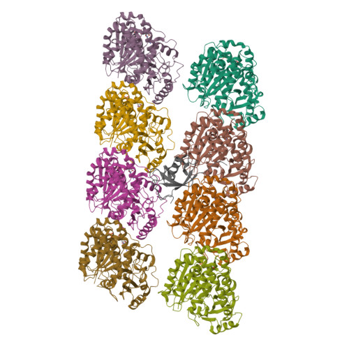
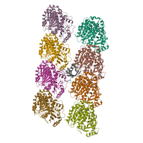
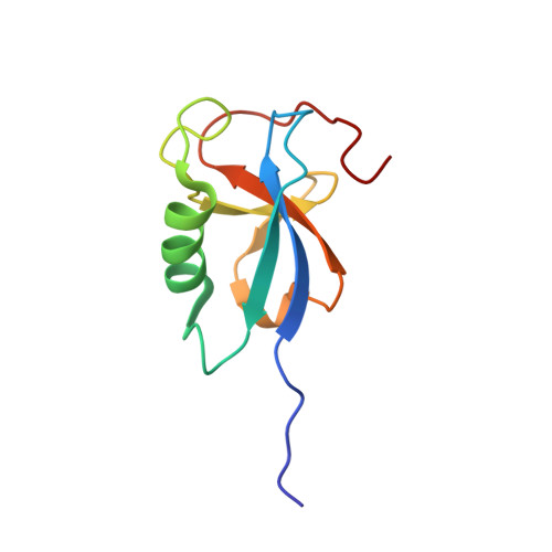
2XRP

Human Doublecortin N-DC Repeat (1MJD) and Mammalian Tubulin (1JFF and 3HKE) Docked into the 8-Angstrom Cryo-EM Map of Doublecortin- Stabilised Microtubules


Experimental Data Snapshot

  • Method: ELECTRON MICROSCOPY
  • Resolution: 8.20 Å
  • Aggregation State: FILAMENT 
  • Reconstruction Method: SINGLE PARTICLE 

Starting Models: experimental
View more details

wwPDB Validation   3D Report Full Report


This is version 2.1 of the entry. See complete history


Literature

Template-Free 13-Protofilament Microtubule-Map Assembly Visualized at 8 A Resolution.

Fourniol, F.J.Sindelar, C.V.Amigues, B.Clare, D.K.Thomas, G.Perderiset, M.Francis, F.Houdusse, A.Moores, C.A.

(2010) J Cell Biol 191: 463

  • DOI: https://doi.org/10.1083/jcb.201007081
  • Primary Citation of Related Structures:  
    2XRP

  • PubMed Abstract: 

    Microtubule-associated proteins (MAPs) are essential for regulating and organizing cellular microtubules (MTs). However, our mechanistic understanding of MAP function is limited by a lack of detailed structural information. Using cryo-electron microscopy and single particle algorithms, we solved the 8 Å structure of doublecortin (DCX)-stabilized MTs. Because of DCX's unusual ability to specifically nucleate and stabilize 13-protofilament MTs, our reconstruction provides unprecedented insight into the structure of MTs with an in vivo architecture, and in the absence of a stabilizing drug. DCX specifically recognizes the corner of four tubulin dimers, a binding mode ideally suited to stabilizing both lateral and longitudinal lattice contacts. A striking consequence of this is that DCX does not bind the MT seam. DCX binding on the MT surface indirectly stabilizes conserved tubulin-tubulin lateral contacts in the MT lumen, operating independently of the nucleotide bound to tubulin. DCX's exquisite binding selectivity uncovers important insights into regulation of cellular MTs.


  • Organizational Affiliation

    Institute of Structural and Molecular Biology, Birkbeck College, London, England, UK.


Macromolecules
Find similar proteins by:  (by identity cutoff)  |  3D Structure
Entity ID: 1
MoleculeChains Sequence LengthOrganismDetailsImage
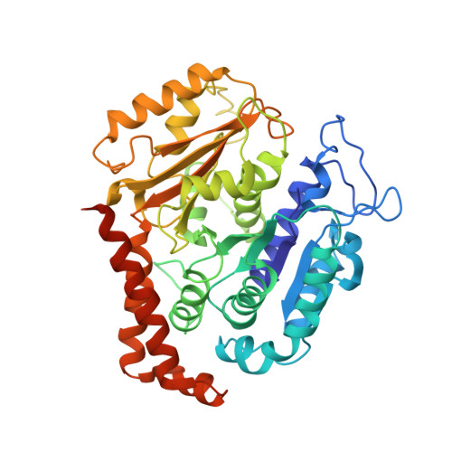
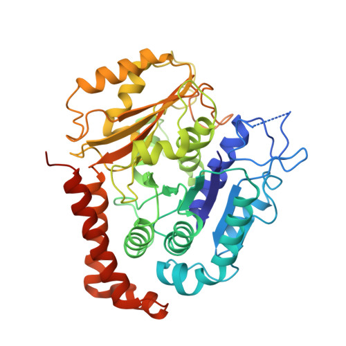
TUBULIN BETA-2B CHAIN
A, C, E, G
445Bos taurusMutation(s): 0 
EC: 3.6.5.6
UniProt
Find proteins for Q6B856 (Bos taurus)
Explore Q6B856 
Go to UniProtKB:  Q6B856
Entity Groups  
Sequence Clusters30% Identity50% Identity70% Identity90% Identity95% Identity100% Identity
UniProt GroupQ6B856
Sequence Annotations
Expand
  • Reference Sequence
Find similar proteins by:  (by identity cutoff)  |  3D Structure
Entity ID: 2
MoleculeChains Sequence LengthOrganismDetailsImage
TUBULIN ALPHA-1D CHAIN
B, D, F, H
452Bos taurusMutation(s): 0 
EC: 3.6.5.6 (PDB Primary Data), 3.6.5 (UniProt)
UniProt
Find proteins for Q2HJ86 (Bos taurus)
Explore Q2HJ86 
Go to UniProtKB:  Q2HJ86
Entity Groups  
Sequence Clusters30% Identity50% Identity70% Identity90% Identity95% Identity100% Identity
UniProt GroupQ2HJ86
Sequence Annotations
Expand
  • Reference Sequence
Find similar proteins by:  (by identity cutoff)  |  3D Structure
Entity ID: 3
MoleculeChains Sequence LengthOrganismDetailsImage
NEURONAL MIGRATION PROTEIN DOUBLECORTIN95Homo sapiensMutation(s): 0 
UniProt & NIH Common Fund Data Resources
Find proteins for O43602 (Homo sapiens)
Explore O43602 
Go to UniProtKB:  O43602
PHAROS:  O43602
GTEx:  ENSG00000077279 
Entity Groups  
Sequence Clusters30% Identity50% Identity70% Identity90% Identity95% Identity100% Identity
UniProt GroupO43602
Sequence Annotations
Expand
  • Reference Sequence
Small Molecules
Ligands 2 Unique
IDChains Name / Formula / InChI Key2D Diagram3D Interactions
GTP
Query on GTP

Download Ideal Coordinates CCD File 
K [auth B],
M [auth D],
O [auth F],
Q [auth H]
GUANOSINE-5'-TRIPHOSPHATE
C10 H16 N5 O14 P3
XKMLYUALXHKNFT-UUOKFMHZSA-N
GDP
Query on GDP

Download Ideal Coordinates CCD File 
J [auth A],
L [auth C],
N [auth E],
P [auth G]
GUANOSINE-5'-DIPHOSPHATE
C10 H15 N5 O11 P2
QGWNDRXFNXRZMB-UUOKFMHZSA-N
Experimental Data & Validation

Experimental Data

  • Method: ELECTRON MICROSCOPY
  • Resolution: 8.20 Å
  • Aggregation State: FILAMENT 
  • Reconstruction Method: SINGLE PARTICLE 
EM Software:
TaskSoftware PackageVersion
RECONSTRUCTIONFREALIGN
RECONSTRUCTIONSPIDER

Structure Validation

View Full Validation Report


Entry History 

Deposition Data

Revision History  (Full details and data files)

  • Version 1.0: 2010-11-24
    Type: Initial release
  • Version 1.1: 2012-03-21
    Changes: Other, Version format compliance
  • Version 2.0: 2017-08-23
    Changes: Atomic model, Data collection, Refinement description
  • Version 2.1: 2024-05-08
    Changes: Data collection, Database references, Derived calculations, Refinement description