2XPP

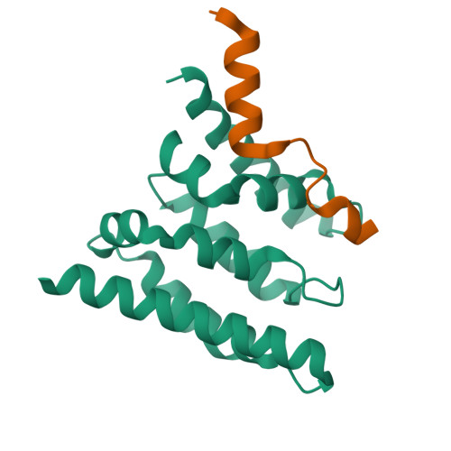
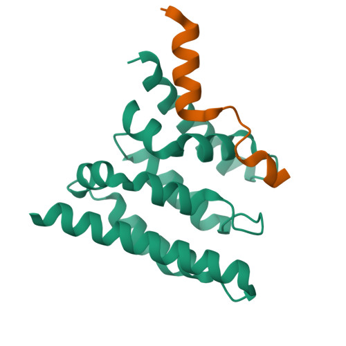
Crystal structure of a Spt6-Iws1(Spn1) complex from Encephalitozoon cuniculi, Form III


Experimental Data Snapshot

  • Method: X-RAY DIFFRACTION
  • Resolution: 1.74 Å
  • R-Value Free: 0.234 
  • R-Value Work: 0.194 
  • R-Value Observed: 0.196 

Starting Model: experimental
View more details

wwPDB Validation   3D Report Full Report


This is version 1.2 of the entry. See complete history


Literature

The Structure of an Iws1/Spt6 Complex Reveals an Interaction Domain Conserved in Tfiis, Elongin a and Med26

Diebold, M.-L.Koch, M.Loeliger, E.Cura, V.Winston, F.Cavarelli, J.Romier, C.

(2010) EMBO J 29: 3979

  • DOI: https://doi.org/10.1038/emboj.2010.272
  • Primary Citation of Related Structures:  
    2XPL, 2XPN, 2XPO, 2XPP

  • PubMed Abstract: 

    Binding of elongation factor Spt6 to Iws1 provides an effective means for coupling eukaryotic mRNA synthesis, chromatin remodelling and mRNA export. We show that an N-terminal region of Spt6 (Spt6N) is responsible for interaction with Iws1. The crystallographic structures of Encephalitozoon cuniculi Iws1 and the Iws1/Spt6N complex reveal two conserved binding subdomains in Iws1. The first subdomain (one HEAT repeat; HEAT subdomain) is a putative phosphoprotein-binding site most likely involved in an Spt6-independent function of Iws1. The second subdomain (two ARM repeats; ARM subdomain) specifically recognizes a bipartite N-terminal region of Spt6. Mutations that alter this region of Spt6 cause severe phenotypes in vivo. Importantly, the ARM subdomain of Iws1 is conserved in several transcription factors, including TFIIS, Elongin A and Med26. We show that the homologous region in yeast TFIIS enables this factor to interact with SAGA and the Mediator subunits Spt8 and Med13, suggesting the molecular basis for TFIIS recruitment at promoters. Taken together, our results provide new structural information about the Iws1/Spt6 complex and reveal a novel interaction domain used for the formation of transcription networks.


  • Organizational Affiliation

    Département de Biologie et Génomique Structurales, IGBMC (Institut de Génétique et Biologie Moléculaire et Cellulaire), UDS, CNRS, INSERM, Illkirch Cedex, France.


Macromolecules
Find similar proteins by:  (by identity cutoff)  |  3D Structure
Entity ID: 1
MoleculeChains Sequence LengthOrganismDetailsImage
IWS1145Encephalitozoon cuniculiMutation(s): 0 
UniProt
Find proteins for Q8SUS7 (Encephalitozoon cuniculi (strain GB-M1))
Explore Q8SUS7 
Go to UniProtKB:  Q8SUS7
Entity Groups  
Sequence Clusters30% Identity50% Identity70% Identity90% Identity95% Identity100% Identity
UniProt GroupQ8SUS7
Sequence Annotations
Expand
  • Reference Sequence
Find similar proteins by:  (by identity cutoff)  |  3D Structure
Entity ID: 2
MoleculeChains Sequence LengthOrganismDetailsImage
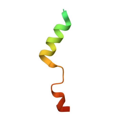
CHROMATIN STRUCTURE MODULATOR42Encephalitozoon cuniculiMutation(s): 0 
UniProt
Find proteins for Q8SRG7 (Encephalitozoon cuniculi (strain GB-M1))
Explore Q8SRG7 
Go to UniProtKB:  Q8SRG7
Entity Groups  
Sequence Clusters30% Identity50% Identity70% Identity90% Identity95% Identity100% Identity
UniProt GroupQ8SRG7
Sequence Annotations
Expand
  • Reference Sequence
Experimental Data & Validation

Experimental Data

  • Method: X-RAY DIFFRACTION
  • Resolution: 1.74 Å
  • R-Value Free: 0.234 
  • R-Value Work: 0.194 
  • R-Value Observed: 0.196 
  • Space Group: P 21 21 21
Unit Cell:
Length ( Å )Angle ( ˚ )
a = 27.356α = 90
b = 56.053β = 90
c = 116.444γ = 90
Software Package:
Software NamePurpose
REFMACrefinement
DENZOdata reduction
SCALEPACKdata scaling
PHASERphasing

Structure Validation

View Full Validation Report


Entry History 

Deposition Data

Revision History  (Full details and data files)

  • Version 1.0: 2010-11-17
    Type: Initial release
  • Version 1.1: 2011-09-21
    Changes: Database references, Version format compliance
  • Version 1.2: 2023-12-20
    Changes: Data collection, Database references, Other, Refinement description