2XPK | pdb_00002xpk

Cell-penetrant, nanomolar O-GlcNAcase inhibitors selective against lysosomal hexosaminidases


Experimental Data Snapshot

  • Method: X-RAY DIFFRACTION
  • Resolution: 2.40 Å
  • R-Value Free: 
    0.242 (Depositor), 0.234 (DCC) 
  • R-Value Work: 
    0.196 (Depositor), 0.192 (DCC) 
  • R-Value Observed: 
    0.196 (Depositor) 

Starting Model: experimental
View more details

wwPDB Validation 3D Report Full Report

Validation slider image for 2XPK

Ligand Structure Quality Assessment 


This is version 1.3 of the entry. See complete history

Literature

Cell-Penetrant, Nanomolar O-Glcnacase Inhibitors Selective Against Lysosomal Hexosaminidases.

Dorfmueller, H.C.Borodkin, V.S.Schimpl, M.Zheng, X.Kime, R.Read, K.D.Van Aalten, D.M.F.

(2010) Chem Biol 17: 1250

  • DOI: https://doi.org/10.1016/j.chembiol.2010.09.014
  • Primary Citation Related Structures: 
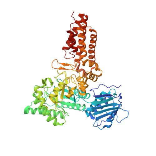
    2XPK

  • PubMed Abstract: 

    Posttranslational modification of metazoan nucleocytoplasmic proteins with N-acetylglucosamine (O-GlcNAc) is essential, dynamic, and inducible and can compete with protein phosphorylation in signal transduction. Inhibitors of O-GlcNAcase, the enzyme removing O-GlcNAc, are useful tools for studying the role of O-GlcNAc in a range of cellular processes. We report the discovery of nanomolar OGA inhibitors that are up to 900,000-fold selective over the related lysosomal hexosaminidases. When applied at nanomolar concentrations on live cells, these cell-penetrant molecules shift the O-GlcNAc equilibrium toward hyper-O-GlcNAcylation with EC₅₀ values down to 3 nM and are thus invaluable tools for the study of O-GlcNAc cell biology.


  • Organizational Affiliation
    • Division of Molecular Microbiology, College of Life Sciences, University of Dundee, Dundee DD15EH, Scotland.

Macromolecule Content 

  • Total Structure Weight: 134.17 kDa 
  • Atom Count: 9,771 
  • Modeled Residue Count: 1,170 
  • Deposited Residue Count: 1,188 
  • Unique protein chains: 1

Macromolecules

Find similar proteins by:|  3D Structure
Entity ID: 1
MoleculeChains  Sequence LengthOrganismDetailsImage
O-GLCNACASE NAGJ
A, B
594Clostridium perfringensMutation(s): 1 
EC: 3.2.1.52 (PDB Primary Data), 3.2.1.169 (UniProt)
UniProt
Find proteins for Q0TR53 (Clostridium perfringens (strain ATCC 13124 / DSM 756 / JCM 1290 / NCIMB 6125 / NCTC 8237 / Type A))
Explore Q0TR53 
Go to UniProtKB:  Q0TR53
Entity Groups
Sequence Clusters30% Identity50% Identity70% Identity90% Identity95% Identity100% Identity
UniProt GroupQ0TR53
Sequence Annotations
Expand
Reference Sequence

Small Molecules

Ligands 1 Unique
IDChains Name / Formula / InChI Key2D Diagram3D Interactions
Z0M

Query on Z0M


Download:Ideal Coordinates CCD File
C [auth A],
D [auth B]
N-[(5R,6R,7R,8S)-6,7-DIHYDROXY-5-(HYDROXYMETHYL)-2-(2-PHENYLETHYL)-5,6,7,8-TETRAHYDROIMIDAZO[1,2-A]PYRIDIN-8-YL]-3-SULFANYLPROPANAMIDE
C19 H25 N3 O4 S
ZEEDRHPJCAOZKV-VDHUWJSZSA-N

Experimental Data & Validation

Experimental Data

  • Method: X-RAY DIFFRACTION
  • Resolution: 2.40 Å
  • R-Value Free:  0.242 (Depositor), 0.234 (DCC) 
  • R-Value Work:  0.196 (Depositor), 0.192 (DCC) 
  • R-Value Observed: 0.196 (Depositor) 
Space Group: I 21 21 21
Unit Cell:
Length ( Å )Angle ( ˚ )
a = 130.145α = 90
b = 144.97β = 90
c = 153.061γ = 90
Software Package:
Software NamePurpose
REFMACrefinement
MOSFLMdata reduction
SCALEPACKdata scaling
REFMACphasing

Structure Validation

View Full Validation Report


Ligand Structure Quality Assessment 


Entry History 

Deposition Data

Revision History  (Full details and data files)

  • Version 1.0: 2011-03-16
    Type: Initial release
  • Version 1.1: 2011-05-08
    Changes: Version format compliance
  • Version 1.2: 2011-07-13
    Changes: Version format compliance
  • Version 1.3: 2023-12-20
    Changes: Data collection, Database references, Derived calculations, Other, Refinement description