2XOI | pdb_00002xoi

Functional and Structural Analyses of N-Acylsulfonamide-Linked Dinucleoside Inhibitors of Ribonuclease A


Experimental Data Snapshot

  • Method: X-RAY DIFFRACTION
  • Resolution: 1.72 Å
  • R-Value Free: 
    0.246 (Depositor) 
  • R-Value Work: 
    0.215 (Depositor), 0.211 (DCC) 
  • R-Value Observed: 
    0.217 (Depositor) 

Starting Model: experimental
View more details

wwPDB Validation 3D Report Full Report

Validation slider image for 2XOI

Ligand Structure Quality Assessment 


This is version 1.4 of the entry. See complete history

Literature

Functional and Structural Analyses of N-Acylsulfonamide-Linked Dinucleoside Inhibitors of Ribonuclease A.

Thiyagarajan, N.Smith, B.D.Raines, R.T.Ravi Acharya, K.

(2011) FEBS J 278: 541

  • DOI: https://doi.org/10.1111/j.1742-4658.2010.07976.x
  • Primary Citation Related Structures: 
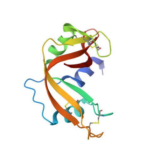
    2XOG, 2XOI

  • PubMed Abstract: 

    Molecular probes are useful for both studying and controlling the functions of enzymes and other proteins. The most useful probes have high affinity for their target, along with small size and resistance to degradation. Here, we report on new surrogates for nucleic acids that fulfill these criteria. Isosteres in which phosphoryl [R-O-P(O(2)(-))-O-R'] groups are replaced with N-acylsulfonamidyl [R-C(O)-N(-)-S(O(2))-R'] or sulfonimidyl [R-S(O(2))-N(-)-S(O(2))-R'] groups increase the number of nonbridging oxygens from two (phosphoryl) to three (N-acylsulfonamidyl) or four (sulfonimidyl). Six such isosteres were found to be more potent inhibitors of catalysis by bovine pancreatic RNase A than are parent compounds containing phosphoryl groups. The atomic structures of two RNase A·N-acylsulfonamide complexes were determined at high resolution by X-ray crystallography. The N-acylsulfonamidyl groups were observed to form more hydrogen bonds with active site residues than did the phosphoryl groups in analogous complexes. These data encourage the further development and use of N-acylsulfonamides and sulfonimides as antagonists of nucleic acid-binding proteins.


  • Organizational Affiliation
    • Department of Biology and Biochemistry, University of Bath, UK.

Macromolecule Content 

  • Total Structure Weight: 28.59 kDa 
  • Atom Count: 2,209 
  • Modeled Residue Count: 245 
  • Deposited Residue Count: 248 
  • Unique protein chains: 1

Macromolecules

Find similar proteins by:|  3D Structure
Entity ID: 1
MoleculeChains  Sequence LengthOrganismDetailsImage
RIBONUCLEASE PANCREATIC
A, B
124Bos taurusMutation(s): 0 
EC: 3.1.27.5 (PDB Primary Data), 4.6.1.18 (UniProt)
UniProt
Find proteins for P61823 (Bos taurus)
Explore P61823 
Go to UniProtKB:  P61823
Entity Groups
Sequence Clusters30% Identity50% Identity70% Identity90% Identity95% Identity100% Identity
UniProt GroupP61823
Sequence Annotations
Expand
Reference Sequence

Small Molecules

Ligands 1 Unique
IDChains Name / Formula / InChI Key2D Diagram3D Interactions
SFB

Query on SFB


Download:Ideal Coordinates CCD File
C [auth A],
D [auth B]
(2S,3S,4R,5R)-5-(6-AMINOPURIN-9-YL)-N-[[(2S,3S,4R,5R)-5-(2,4-DIOXOPYRIMIDIN-1-YL)-4-HYDROXY-2-(HYDROXYMETHYL)OXOLAN-3-YL]METHYLSULFONYL]-3,4-DIHYDROXY-OXOLANE-2-CARBOXAMIDE
C20 H28 N8 O11 S
KEQMCGJHYDWPEW-ZSIUBJGCSA-N

Experimental Data & Validation

Experimental Data

  • Method: X-RAY DIFFRACTION
  • Resolution: 1.72 Å
  • R-Value Free:  0.246 (Depositor) 
  • R-Value Work:  0.215 (Depositor), 0.211 (DCC) 
  • R-Value Observed: 0.217 (Depositor) 
Space Group: C 1 2 1
Unit Cell:
Length ( Å )Angle ( ˚ )
a = 100.787α = 90
b = 33.146β = 90.94
c = 72.748γ = 90
Software Package:
Software NamePurpose
REFMACrefinement
HKL-2000data reduction
HKL-2000data scaling
MOLREPphasing

Structure Validation

View Full Validation Report


Ligand Structure Quality Assessment 


Entry History 

Deposition Data

Revision History  (Full details and data files)

  • Version 1.0: 2011-01-19
    Type: Initial release
  • Version 1.1: 2011-05-08
    Changes: Version format compliance
  • Version 1.2: 2011-07-13
    Changes: Version format compliance
  • Version 1.3: 2023-12-20
    Changes: Data collection, Database references, Derived calculations, Other, Refinement description
  • Version 1.4: 2024-11-06
    Changes: Structure summary