2XNS

Crystal Structure Of Human G alpha i1 Bound To A Designed Helical Peptide Derived From The Goloco Motif Of RGS14


Experimental Data Snapshot

  • Method: X-RAY DIFFRACTION
  • Resolution: 3.41 Å
  • R-Value Free: 0.244 
  • R-Value Work: 0.223 
  • R-Value Observed: 0.223 

Starting Model: experimental
View more details

wwPDB Validation   3D Report Full Report


Ligand Structure Quality Assessment 


This is version 1.3 of the entry. See complete history


Literature

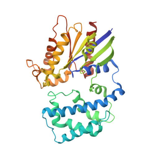
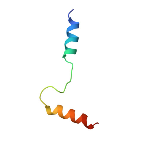
Computational Design of the Sequence and Structure of a Protein-Binding Peptide.

Sammond, D.W.Bosch, D.E.Butterfoss, G.L.Purbeck, C.Machius, M.Siderovski, D.P.Kuhlman, B.

(2011) J Am Chem Soc 133: 4190

  • DOI: https://doi.org/10.1021/ja110296z
  • Primary Citation of Related Structures:  
    2XNS

  • PubMed Abstract: 

    The de novo design of protein-binding peptides is challenging because it requires the identification of both a sequence and a backbone conformation favorable for binding. We used a computational strategy that iterates between structure and sequence optimization to redesign the C-terminal portion of the RGS14 GoLoco motif peptide so that it adopts a new conformation when bound to Gα(i1). An X-ray crystal structure of the redesigned complex closely matches the computational model, with a backbone root-mean-square deviation of 1.1 Å.


  • Organizational Affiliation

    Department of Biochemistry and Biophysics, University of North Carolina, Chapel Hill, North Carolina 27599-7260, USA.


Macromolecules
Find similar proteins by:  (by identity cutoff)  |  3D Structure
Entity ID: 1
MoleculeChains Sequence LengthOrganismDetailsImage
GUANINE NUCLEOTIDE-BINDING PROTEIN G(I) SUBUNIT ALPHA-1
A, B
327Homo sapiensMutation(s): 0 
EC: 3.6.5.1
UniProt & NIH Common Fund Data Resources
Find proteins for P63096 (Homo sapiens)
Explore P63096 
Go to UniProtKB:  P63096
PHAROS:  P63096
GTEx:  ENSG00000127955 
Entity Groups  
Sequence Clusters30% Identity50% Identity70% Identity90% Identity95% Identity100% Identity
UniProt GroupP63096
Sequence Annotations
Expand
  • Reference Sequence
Find similar proteins by:  (by identity cutoff)  |  3D Structure
Entity ID: 2
MoleculeChains Sequence LengthOrganismDetailsImage
REGULATOR OF G-PROTEIN SIGNALING 14
C, D
40Homo sapiensMutation(s): 0 
UniProt & NIH Common Fund Data Resources
Find proteins for O43566 (Homo sapiens)
Explore O43566 
Go to UniProtKB:  O43566
PHAROS:  O43566
GTEx:  ENSG00000169220 
Entity Groups  
Sequence Clusters30% Identity50% Identity70% Identity90% Identity95% Identity100% Identity
UniProt GroupO43566
Sequence Annotations
Expand
  • Reference Sequence
Experimental Data & Validation

Experimental Data

  • Method: X-RAY DIFFRACTION
  • Resolution: 3.41 Å
  • R-Value Free: 0.244 
  • R-Value Work: 0.223 
  • R-Value Observed: 0.223 
  • Space Group: I 2 3
Unit Cell:
Length ( Å )Angle ( ˚ )
a = 265.261α = 90
b = 265.261β = 90
c = 265.261γ = 90
Software Package:
Software NamePurpose
REFMACrefinement
HKL-2000data reduction
HKL-2000data scaling
PHASERphasing

Structure Validation

View Full Validation Report


Ligand Structure Quality Assessment 


Entry History 

Deposition Data

Revision History  (Full details and data files)

  • Version 1.0: 2011-06-08
    Type: Initial release
  • Version 1.1: 2011-07-13
    Changes: Version format compliance
  • Version 1.2: 2019-05-08
    Changes: Data collection, Experimental preparation, Other
  • Version 1.3: 2023-12-20
    Changes: Data collection, Database references, Derived calculations, Other, Refinement description