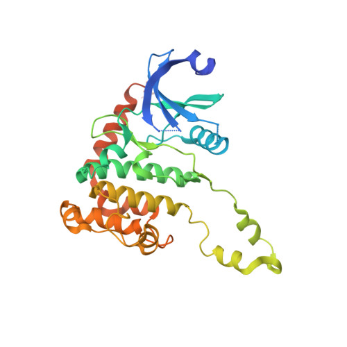
2XM9

Structure of a small molecule inhibitor with the kinase domain of Chk2


Experimental Data Snapshot

  • Method: X-RAY DIFFRACTION
  • Resolution: 2.50 Å
  • R-Value Free: 0.238 
  • R-Value Work: 0.186 
  • R-Value Observed: 0.189 

Starting Model: experimental
View more details

wwPDB Validation   3D Report Full Report


Ligand Structure Quality Assessment 


This is version 1.3 of the entry. See complete history


Literature

Structure-Based Design of Potent and Selective 2-(Quinazolin-2-Yl)Phenol Inhibitors of Checkpoint Kinase 2.

Caldwell, J.J.Welsh, E.J.Matijssen, C.Anderson, V.E.Antoni, L.Boxall, K.Urban, F.Hayes, A.Raynaud, F.I.Rigoreau, L.J.Raynham, T.Aherne, G.W.Pearl, L.H.Oliver, A.W.Garrett, M.D.Collins, I.

(2011) J Med Chem 54: 580

  • DOI: https://doi.org/10.1021/jm101150b
  • Primary Citation of Related Structures:  
    2XBJ, 2XM8, 2XM9

  • PubMed Abstract: 

    Structure-based design was applied to the optimization of a series of 2-(quinazolin-2-yl)phenols to generate potent and selective ATP-competitive inhibitors of the DNA damage response signaling enzyme checkpoint kinase 2 (CHK2). Structure-activity relationships for multiple substituent positions were optimized separately and in combination leading to the 2-(quinazolin-2-yl)phenol 46 (IC(50) 3 nM) with good selectivity for CHK2 against CHK1 and a wider panel of kinases and with promising in vitro ADMET properties. Off-target activity at hERG ion channels shown by the core scaffold was successfully reduced by the addition of peripheral polar substitution. In addition to showing mechanistic inhibition of CHK2 in HT29 human colon cancer cells, a concentration dependent radioprotective effect in mouse thymocytes was demonstrated for the potent inhibitor 46 (CCT241533).


  • Organizational Affiliation

    Cancer Research UK Cancer Therapeutics Unit, The Institute of Cancer Research, 15 Cotswold Road, Sutton, Surrey SM2 5NG, UK. john.caldwell@icr.ac.uk


Macromolecules
Find similar proteins by:  (by identity cutoff)  |  3D Structure
Entity ID: 1
MoleculeChains Sequence LengthOrganismDetailsImage
SERINE/THREONINE-PROTEIN KINASE CHK2329Homo sapiensMutation(s): 0 
EC: 2.7.11.1
UniProt & NIH Common Fund Data Resources
Find proteins for O96017 (Homo sapiens)
Explore O96017 
Go to UniProtKB:  O96017
PHAROS:  O96017
GTEx:  ENSG00000183765 
Entity Groups  
Sequence Clusters30% Identity50% Identity70% Identity90% Identity95% Identity100% Identity
UniProt GroupO96017
Sequence Annotations
Expand
  • Reference Sequence
Small Molecules
Ligands 2 Unique
IDChains Name / Formula / InChI Key2D Diagram3D Interactions
LWH
Query on LWH

Download Ideal Coordinates CCD File 
B [auth A]4-(1H-pyrazol-5-yl)-2-{4-[(3S)-pyrrolidin-3-ylamino]quinazolin-2-yl}phenol
C21 H20 N6 O
DZNFGQDAMCNMLC-AWEZNQCLSA-N
NO3
Query on NO3

Download Ideal Coordinates CCD File 
C [auth A]NITRATE ION
N O3
NHNBFGGVMKEFGY-UHFFFAOYSA-N
Binding Affinity Annotations 
IDSourceBinding Affinity
LWH PDBBind:  2XM9 IC50: 9 (nM) from 1 assay(s)
Experimental Data & Validation

Experimental Data

  • Method: X-RAY DIFFRACTION
  • Resolution: 2.50 Å
  • R-Value Free: 0.238 
  • R-Value Work: 0.186 
  • R-Value Observed: 0.189 
  • Space Group: P 32 2 1
Unit Cell:
Length ( Å )Angle ( ˚ )
a = 91.36α = 90
b = 91.36β = 90
c = 92.95γ = 120
Software Package:
Software NamePurpose
PHENIXrefinement
iMOSFLMdata reduction
SCALAdata scaling
PHASERphasing

Structure Validation

View Full Validation Report


Ligand Structure Quality Assessment 


Entry History 

Revision History  (Full details and data files)

  • Version 1.0: 2011-01-12
    Type: Initial release
  • Version 1.1: 2011-05-08
    Changes: Version format compliance
  • Version 1.2: 2011-07-13
    Changes: Version format compliance
  • Version 1.3: 2023-12-20
    Changes: Data collection, Database references, Derived calculations, Other, Refinement description