2XLL | pdb_00002xll

The crystal structure of bilirubin oxidase from Myrothecium verrucaria


Experimental Data Snapshot

  • Method: X-RAY DIFFRACTION
  • Resolution: 2.31 Å
  • R-Value Free: 
    0.216 (Depositor), 0.209 (DCC) 
  • R-Value Work: 
    0.170 (Depositor), 0.170 (DCC) 
  • R-Value Observed: 
    0.172 (Depositor) 

Starting Model: experimental
View more details

wwPDB Validation 3D Report Full Report

Validation slider image for 2XLL

This is version 2.2 of the entry. See complete history

Literature

Bilirubin Oxidase from Myrothecium Verrucaria: X- Ray Determination of the Complete Crystal Structure and a Rational Surface Modification for Enhanced Electrocatalytic O(2) Reduction.

Cracknell, J.A.Mcnamara, T.P.Lowe, E.D.Blanford, C.F.

(2011) Dalton Trans 40: 6668

  • DOI: https://doi.org/10.1039/c0dt01403f
  • Primary Citation Related Structures: 
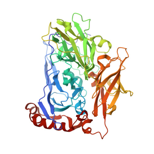
    2XLL

  • PubMed Abstract: 

    The blue multi-copper oxidase bilirubin oxidase (BOx) from the ascomycete plant pathogen Myrothecium verrucaria (Mv) efficiently catalyses the oxidation of bilirubin to biliverdin, with the concomitant reduction of O(2) to water, a reaction of considerable interest for low-temperature bio-fuel cell applications. We have solved the complete X-ray determined structure of Mv BOx at 2.4 Å resolution, using molecular replacement with the Spore Coat Protein A (CotA) enzyme from Bacillus subtilis (PDB code 1GSK) as a template. The structure reveals an unusual environment around the blue type 1 copper (T1 Cu) that includes two non-coordinating hydrophilic amino acids, asparagine and threonine. The presence of a long, narrow and hydrophilic pocket near the T1 Cu suggests that structure of the substrate-binding site is dynamically determined in vivo. We show that the interaction between the binding pocket of Mv BOx and its highly conjugated natural organic substrate, bilirubin, can be used to stabilise the enzyme on a pyrolytic graphite electrode, more than doubling its electrocatalytic activity relative to the current obtained by simple adsorption of the protein to the carbon surface.


  • Organizational Affiliation
    • Department of Chemistry, Inorganic Chemistry Laboratory, University of Oxford, South Parks Road, Oxford, UK OX1 3QR.

Macromolecule Content 

  • Total Structure Weight: 244.45 kDa 
  • Atom Count: 18,363 
  • Modeled Residue Count: 2,132 
  • Deposited Residue Count: 2,136 
  • Unique protein chains: 1

Macromolecules

Find similar proteins by:|  3D Structure
Entity ID: 1
MoleculeChains  Sequence LengthOrganismDetailsImage
BILIRUBIN OXIDASE
A, B, C, D
534Albifimbria verrucariaMutation(s): 0 
EC: 1.3.3.5
UniProt
Find proteins for Q12737 (Albifimbria verrucaria)
Explore Q12737 
Go to UniProtKB:  Q12737
Entity Groups
Sequence Clusters30% Identity50% Identity70% Identity90% Identity95% Identity100% Identity
UniProt GroupQ12737
Glycosylation
Glycosylation Sites: 2
Sequence Annotations
Expand
Reference Sequence

Oligosaccharides

Help  
Entity ID: 2
MoleculeChains Length2D Diagram GlycosylationD Interactions
2-acetamido-2-deoxy-beta-D-glucopyranose-(1-4)-2-acetamido-2-deoxy-beta-D-glucopyranose
E, F, G, H, I
E, F, G, H, I, J, K, L
2N-Glycosylation
Glycosylation Resources
GlyTouCan: G42666HT
GlyCosmos: G42666HT
GlyGen: G42666HT

Small Molecules

Ligands 1 Unique
IDChains Name / Formula / InChI Key2D Diagram3D Interactions
CU

Query on CU


Download:Ideal Coordinates CCD File
AA [auth D]
BA [auth D]
M [auth A]
N [auth A]
O [auth A]
AA [auth D],
BA [auth D],
M [auth A],
N [auth A],
O [auth A],
P [auth A],
Q [auth B],
R [auth B],
S [auth B],
T [auth B],
U [auth C],
V [auth C],
W [auth C],
X [auth C],
Y [auth D],
Z [auth D]
COPPER (II) ION
Cu
JPVYNHNXODAKFH-UHFFFAOYSA-N

Experimental Data & Validation

Experimental Data

  • Method: X-RAY DIFFRACTION
  • Resolution: 2.31 Å
  • R-Value Free:  0.216 (Depositor), 0.209 (DCC) 
  • R-Value Work:  0.170 (Depositor), 0.170 (DCC) 
  • R-Value Observed: 0.172 (Depositor) 
Space Group: P 1
Unit Cell:
Length ( Å )Angle ( ˚ )
a = 52.793α = 89.98
b = 83.602β = 89.89
c = 143.148γ = 89.9
Software Package:
Software NamePurpose
PHENIXrefinement
XDSdata reduction
SCALAdata scaling
PHASERphasing

Structure Validation

View Full Validation Report


Entry History 

Deposition Data

Revision History  (Full details and data files)

  • Version 1.0: 2011-04-20
    Type: Initial release
  • Version 1.1: 2011-06-23
    Changes: Version format compliance
  • Version 1.2: 2011-07-13
    Changes: Version format compliance
  • Version 2.0: 2020-07-29
    Type: Remediation
    Reason: Carbohydrate remediation
    Changes: Advisory, Atomic model, Data collection, Derived calculations, Other, Structure summary
  • Version 2.1: 2023-12-20
    Changes: Advisory, Data collection, Database references, Refinement description, Structure summary
  • Version 2.2: 2024-11-06
    Changes: Structure summary