2XJM | pdb_00002xjm

Crystal structure of Streptococcus suis Dpr with cobalt


Experimental Data Snapshot

  • Method: X-RAY DIFFRACTION
  • Resolution: 2.30 Å
  • R-Value Free: 
    0.240 (Depositor), 0.247 (DCC) 
  • R-Value Work: 
    0.177 (Depositor), 0.190 (DCC) 
  • R-Value Observed: 
    0.180 (Depositor) 

wwPDB Validation 3D Report Full Report

Validation slider image for 2XJM

This is version 1.3 of the entry. See complete history

Literature

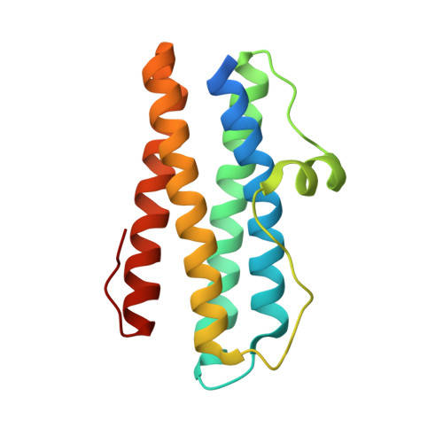
Structural and Thermodynamic Characterization of Metal Ion Binding in Streptococcus Suis Dpr.

Haikarainen, T.Thanassoulas, A.Stavros, P.Nounesis, G.Haataja, S.Papageorgiou, A.C.

(2011) J Mol Biology 405: 448

  • DOI: https://doi.org/10.1016/j.jmb.2010.10.058
  • Primary Citation Related Structures: 
    2XJM, 2XJN, 2XJO, 2XKQ

  • PubMed Abstract: 

    The use of protein cages for the creation of novel inorganic nanomaterials has attracted considerable attention in recent years. Ferritins are among the most commonly used protein cages in nanoscience. Accordingly, the binding of various metals to ferritins has been studied extensively. Dps (DNA-binding protein from starved cells)-like proteins belong to the ferritin superfamily. In contrast to ferritins, Dps-like proteins form 12-mers instead of 24-mers, have a different ferroxidase center, and are able to store a smaller amount of iron atoms in a hollow cavity (up to ∼500, instead of the ∼4500 iron atoms found in ferritins). With the exception of iron, the binding of other metal cations to Dps proteins has not been studied in detail. Here, the binding of six divalent metal ions (Zn(2+), Mn(2+), Ni(2+), Co(2+), Cu(2+), and Mg(2+)) to Streptococcus suisDps-like peroxide resistance protein (SsDpr) was characterized by X-ray crystallography and isothermal titration calorimetry (ITC). All metal cations, except for Mg(2+), were found to bind to the ferroxidase center similarly to Fe(2+), with moderate affinity (binding constants between 0.1×10(5) M(-1) and 5×10(5) M(-1)). The stoichiometry of binding, as deduced by ITC data, suggested the presence of a dication ferroxidase site. No other metal binding sites were identified in the protein. The results presented here demonstrate the ability of SsDpr to bind various metals as substitutes for iron and will help in better understanding protein-metal interactions in the Dps family of proteins as potential metal nanocontainers.


  • Organizational Affiliation
    • Turku Center for Biotechnology, University of Turku and Åbo Akademi University, Turku 20521, Finland.

Macromolecule Content 

  • Total Structure Weight: 225.78 kDa 
  • Atom Count: 15,464 
  • Modeled Residue Count: 1,811 
  • Deposited Residue Count: 1,980 
  • Unique protein chains: 1

Macromolecules

Find similar proteins by:|  3D Structure
Entity ID: 1
MoleculeChains  Sequence LengthOrganismDetailsImage
DNA PROTECTION DURING STARVATION PROTEIN
A, B, C, D, E
A, B, C, D, E, F, G, H, I, J, K, L
165Streptococcus suisMutation(s): 0 
EC: 1.16
UniProt
Find proteins for P0CB53 (Streptococcus suis)
Explore P0CB53 
Go to UniProtKB:  P0CB53
Entity Groups
Sequence Clusters30% Identity50% Identity70% Identity90% Identity95% Identity100% Identity
UniProt GroupP0CB53
Sequence Annotations
Expand
Reference Sequence

Small Molecules

Ligands 4 Unique
IDChains Name / Formula / InChI Key2D Diagram3D Interactions
EPE

Query on EPE


Download:Ideal Coordinates CCD File
CA [auth I],
GA [auth J],
KA [auth K],
T [auth E]
4-(2-HYDROXYETHYL)-1-PIPERAZINE ETHANESULFONIC ACID
C8 H18 N2 O4 S
JKMHFZQWWAIEOD-UHFFFAOYSA-N
CO

Query on CO


Download:Ideal Coordinates CCD File
AA [auth H]
DA [auth I]
HA [auth J]
LA [auth K]
M [auth A]
AA [auth H],
DA [auth I],
HA [auth J],
LA [auth K],
M [auth A],
MA [auth L],
O [auth B],
Q [auth C],
S [auth D],
U [auth E],
W [auth F],
Z [auth G]
COBALT (II) ION
Co
XLJKHNWPARRRJB-UHFFFAOYSA-N
CA

Query on CA


Download:Ideal Coordinates CCD File
BA [auth H]
FA [auth I]
JA [auth J]
P [auth B]
R [auth C]
BA [auth H],
FA [auth I],
JA [auth J],
P [auth B],
R [auth C],
V [auth E],
X [auth F],
Y [auth F]
CALCIUM ION
Ca
BHPQYMZQTOCNFJ-UHFFFAOYSA-N
CL

Query on CL


Download:Ideal Coordinates CCD File
EA [auth I],
IA [auth J],
N [auth A]
CHLORIDE ION
Cl
VEXZGXHMUGYJMC-UHFFFAOYSA-M

Experimental Data & Validation

Experimental Data

  • Method: X-RAY DIFFRACTION
  • Resolution: 2.30 Å
  • R-Value Free:  0.240 (Depositor), 0.247 (DCC) 
  • R-Value Work:  0.177 (Depositor), 0.190 (DCC) 
  • R-Value Observed: 0.180 (Depositor) 
Space Group: P 21 21 21
Unit Cell:
Length ( Å )Angle ( ˚ )
a = 104.96α = 90
b = 137.42β = 90
c = 141.95γ = 90
Software Package:
Software NamePurpose
REFMACrefinement
XDSdata reduction
XDSdata scaling

Structure Validation

View Full Validation Report


Entry History 

Deposition Data

Revision History  (Full details and data files)

  • Version 1.0: 2010-11-24
    Type: Initial release
  • Version 1.1: 2011-10-12
    Changes: Database references, Version format compliance
  • Version 1.2: 2019-07-24
    Changes: Data collection
  • Version 1.3: 2024-05-08
    Changes: Data collection, Database references, Derived calculations, Other