2XHM | pdb_00002xhm

Crystal structure of AnCE-K26 complex


Experimental Data Snapshot

  • Method: X-RAY DIFFRACTION
  • Resolution: 1.96 Å
  • R-Value Free: 
    0.215 (Depositor), 0.220 (DCC) 
  • R-Value Work: 
    0.194 (Depositor), 0.196 (DCC) 
  • R-Value Observed: 
    0.195 (Depositor) 

Starting Model: experimental
View more details

wwPDB Validation 3D Report Full Report

Validation slider image for 2XHM

Ligand Structure Quality Assessment 


This is version 2.2 of the entry. See complete history

Literature

Crystal Structure of a Phosphonotripeptide K-26 in Complex with Angiotensin Converting Enzyme Homologue (Ance) from Drosophila Melanogaster.

Akif, M.Ntai, I.Sturrock, E.D.Isaac, R.E.Bachmann, B.O.Acharya, K.R.

(2010) Biochem Biophys Res Commun 398: 532

  • DOI: https://doi.org/10.1016/j.bbrc.2010.06.113
  • Primary Citation Related Structures: 
    2XHM

  • PubMed Abstract: 

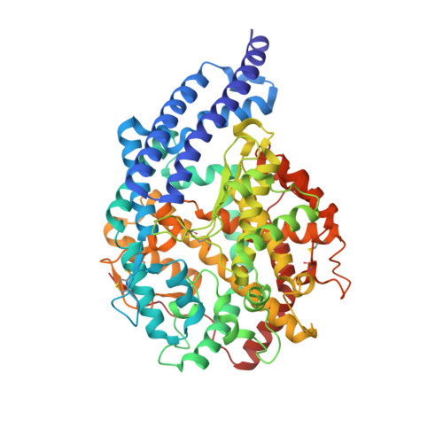
    Angiotensin-I converting enzyme (ACE, a zinc dependent dipeptidyl carboxypeptidase) is a major target of drugs due to its role in the modulation of blood pressure and cardiovascular disorders. Here we present a crystal structure of AnCE (an ACE homologue from Drosophila melanogaster with a single enzymatic domain) in complex with a natural product-phosphonotripeptide, K-26 at 1.96A resolution. The inhibitor binds exclusively in the S(1) and S(2) binding pockets of AnCE (coordinating the zinc ion) through ionic and hydrogen bond interactions. A detailed structural comparison of AnCE.K-26 complex with individual domains of human somatic ACE provides useful information for further exploration of ACE inhibitor pharmacophores involving phosphonic acids.


  • Organizational Affiliation
    • Department of Biology and Biochemistry, University of Bath, Claverton Down, Bath BA2 7AY, UK.

Macromolecule Content 

  • Total Structure Weight: 71.51 kDa 
  • Atom Count: 5,532 
  • Modeled Residue Count: 598 
  • Deposited Residue Count: 598 
  • Unique protein chains: 1

Macromolecules

Find similar proteins by:|  3D Structure
Entity ID: 1
MoleculeChains  Sequence LengthOrganismDetailsImage
ANGIOTENSIN CONVERTING ENZYME598Drosophila melanogasterMutation(s): 0 
EC: 3.4.15.1
UniProt
Find proteins for Q10714 (Drosophila melanogaster)
Explore Q10714 
Go to UniProtKB:  Q10714
Entity Groups
Sequence Clusters30% Identity50% Identity70% Identity90% Identity95% Identity100% Identity
UniProt GroupQ10714
Glycosylation
Glycosylation Sites: 3Go to GlyGen: Q10714-1
Sequence Annotations
Expand
Reference Sequence

Oligosaccharides

Help  
Entity ID: 2
MoleculeChains Length2D Diagram GlycosylationD Interactions
beta-D-mannopyranose-(1-6)-alpha-D-mannopyranose-(1-3)-[alpha-D-mannopyranose-(1-6)]beta-D-mannopyranose-(1-4)-2-acetamido-2-deoxy-beta-D-glucopyranose-(1-4)-2-acetamido-2-deoxy-beta-D-glucopyranose
B
6N-Glycosylation
Glycosylation Resources
GlyTouCan: G64803HZ
GlyCosmos: G64803HZ
GlyGen: G64803HZ

Small Molecules

Ligands 4 Unique
IDChains Name / Formula / InChI Key2D Diagram3D Interactions
K26

Query on K26


Download:Ideal Coordinates CCD File
C [auth A]N-ACETYL-L-ILE-L-TYR-(R)-1-AMINO-2-(4-HYDROXYPHENYL)ETHYLPHOSPHONIC ACID
C25 H34 N3 O8 P
ZFRNBYWFOLDQKG-FDMHNHSTSA-N
EPE

Query on EPE


Download:Ideal Coordinates CCD File
E [auth A]4-(2-HYDROXYETHYL)-1-PIPERAZINE ETHANESULFONIC ACID
C8 H18 N2 O4 S
JKMHFZQWWAIEOD-UHFFFAOYSA-N
NAG

Query on NAG


Download:Ideal Coordinates CCD File
F [auth A],
G [auth A]
2-acetamido-2-deoxy-beta-D-glucopyranose
C8 H15 N O6
OVRNDRQMDRJTHS-FMDGEEDCSA-N
ZN

Query on ZN


Download:Ideal Coordinates CCD File
D [auth A]ZINC ION
Zn
PTFCDOFLOPIGGS-UHFFFAOYSA-N

Experimental Data & Validation

Experimental Data

  • Method: X-RAY DIFFRACTION
  • Resolution: 1.96 Å
  • R-Value Free:  0.215 (Depositor), 0.220 (DCC) 
  • R-Value Work:  0.194 (Depositor), 0.196 (DCC) 
  • R-Value Observed: 0.195 (Depositor) 
Space Group: H 3
Unit Cell:
Length ( Å )Angle ( ˚ )
a = 172.707α = 90
b = 172.707β = 90
c = 101.696γ = 120
Software Package:
Software NamePurpose
REFMACrefinement
HKL-2000data reduction
SCALEPACKdata scaling
PHASERphasing

Structure Validation

View Full Validation Report


Ligand Structure Quality Assessment 


Entry History 

Deposition Data

Revision History  (Full details and data files)

  • Version 1.0: 2010-07-14
    Type: Initial release
  • Version 1.1: 2011-05-08
    Changes: Version format compliance
  • Version 1.2: 2011-07-13
    Changes: Version format compliance
  • Version 2.0: 2020-07-29
    Type: Remediation
    Reason: Carbohydrate remediation
    Changes: Atomic model, Data collection, Derived calculations, Other, Structure summary
  • Version 2.1: 2023-12-20
    Changes: Data collection, Database references, Refinement description, Structure summary
  • Version 2.2: 2024-10-16
    Changes: Structure summary