2XEW | pdb_00002xew

Crystal structure of K11-linked diubiquitin


Experimental Data Snapshot

  • Method: X-RAY DIFFRACTION
  • Resolution: 2.20 Å
  • R-Value Free: 
    0.252 (Depositor), 0.249 (DCC) 
  • R-Value Work: 
    0.205 (Depositor), 0.204 (DCC) 
  • R-Value Observed: 
    0.207 (Depositor) 

Starting Model: experimental
View more details

wwPDB Validation 3D Report Full Report

Validation slider image for 2XEW

This is version 1.4 of the entry. See complete history

Literature

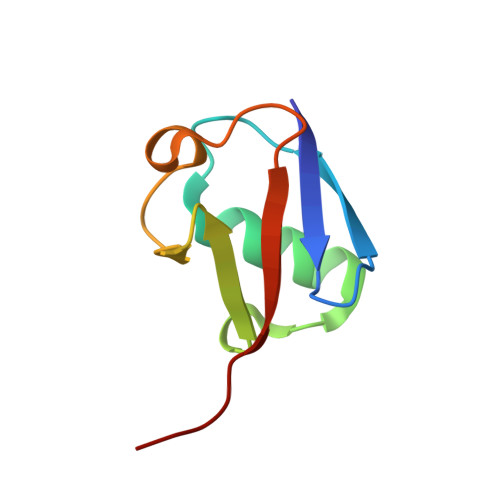
Lys11-Linked Ubiquitin Chains Adopt Compact Conformations and are Preferentially Hydrolyzed by the Deubiquitinase Cezanne

Bremm, A.Freund, S.M.V.Komander, D.

(2010) Nat Struct Mol Biol 17: 939

  • DOI: https://doi.org/10.1038/nsmb.1873
  • Primary Citation Related Structures: 
    2XEW

  • PubMed Abstract: 

    Ubiquitin is a versatile cellular signaling molecule that can form polymers of eight different linkages, and individual linkage types have been associated with distinct cellular functions. Though little is currently known about Lys11-linked ubiquitin chains, recent data indicate that they may be as abundant as Lys48 linkages and may be involved in vital cellular processes. Here we report the generation of Lys11-linked polyubiquitin in vitro, for which the Lys11-specific E2 enzyme UBE2S was fused to a ubiquitin binding domain. Crystallographic and NMR analyses of Lys11-linked diubiquitin reveal that Lys11-linked chains adopt compact conformations in which Ile44 is solvent exposed. Furthermore, we identify the OTU family deubiquitinase Cezanne as the first deubiquitinase with Lys11-linkage preference. Our data highlight the intrinsic specificity of the ubiquitin system that extends to Lys11-linked chains and emphasize that differentially linked polyubiquitin chains must be regarded as independent post-translational modifications.


  • Organizational Affiliation
    • Medical Research Council Laboratory of Molecular Biology, Cambridge, UK.

Macromolecule Content 

  • Total Structure Weight: 104.6 kDa 
  • Atom Count: 7,921 
  • Modeled Residue Count: 908 
  • Deposited Residue Count: 912 
  • Unique protein chains: 1

Macromolecules

Find similar proteins by:|  3D Structure
Entity ID: 1
MoleculeChains  Sequence LengthOrganismDetailsImage
UBIQUITIN
A, B, C, D, E
A, B, C, D, E, F, G, H, I, J, K, L
76Homo sapiensMutation(s): 0 
UniProt & NIH Common Fund Data Resources
Find proteins for P0CG48 (Homo sapiens)
Explore P0CG48 
Go to UniProtKB:  P0CG48
PHAROS:  P0CG48
GTEx:  ENSG00000150991 
Entity Groups
Sequence Clusters30% Identity50% Identity70% Identity90% Identity95% Identity100% Identity
UniProt GroupP0CG48
Sequence Annotations
Expand
Reference Sequence

Small Molecules

Ligands 3 Unique
IDChains Name / Formula / InChI Key2D Diagram3D Interactions
FLC

Query on FLC


Download:Ideal Coordinates CCD File
Y [auth F],
Z [auth F]
CITRATE ANION
C6 H5 O7
KRKNYBCHXYNGOX-UHFFFAOYSA-K
EDO

Query on EDO


Download:Ideal Coordinates CCD File
AA [auth F]
BA [auth H]
CA [auth I]
EA [auth J]
FA [auth K]
AA [auth F],
BA [auth H],
CA [auth I],
EA [auth J],
FA [auth K],
GA [auth L],
HA [auth L],
IA [auth L],
M [auth A],
N [auth A],
O [auth A],
Q [auth B],
R [auth B],
S [auth B],
T [auth C],
V [auth D],
X [auth E]
1,2-ETHANEDIOL
C2 H6 O2
LYCAIKOWRPUZTN-UHFFFAOYSA-N
CL

Query on CL


Download:Ideal Coordinates CCD File
DA [auth I]
JA [auth L]
KA [auth L]
LA [auth L]
P [auth A]
DA [auth I],
JA [auth L],
KA [auth L],
LA [auth L],
P [auth A],
U [auth C],
W [auth D]
CHLORIDE ION
Cl
VEXZGXHMUGYJMC-UHFFFAOYSA-M

Experimental Data & Validation

Experimental Data

  • Method: X-RAY DIFFRACTION
  • Resolution: 2.20 Å
  • R-Value Free:  0.252 (Depositor), 0.249 (DCC) 
  • R-Value Work:  0.205 (Depositor), 0.204 (DCC) 
  • R-Value Observed: 0.207 (Depositor) 
Space Group: P 2 2 21
Unit Cell:
Length ( Å )Angle ( ˚ )
a = 79.23α = 90
b = 79.96β = 90
c = 221.23γ = 90
Software Package:
Software NamePurpose
PHENIXrefinement
iMOSFLMdata reduction
SCALAdata scaling
MOLREPphasing

Structure Validation

View Full Validation Report


Entry History 

Deposition Data

Revision History  (Full details and data files)

  • Version 1.0: 2010-07-14
    Type: Initial release
  • Version 1.1: 2011-05-08
    Changes: Version format compliance
  • Version 1.2: 2011-07-13
    Changes: Version format compliance
  • Version 1.3: 2019-07-31
    Changes: Advisory, Data collection, Derived calculations
  • Version 1.4: 2023-12-20
    Changes: Data collection, Database references, Derived calculations, Other, Refinement description