2XCE | pdb_00002xce

Structure of YncF in complex with dUpNHpp


Experimental Data Snapshot

  • Method: X-RAY DIFFRACTION
  • Resolution: 1.85 Å
  • R-Value Free: 
    0.187 (Depositor), 0.201 (DCC) 
  • R-Value Work: 
    0.151 (Depositor), 0.170 (DCC) 
  • R-Value Observed: 
    0.153 (Depositor) 

Starting Model: experimental
View more details

wwPDB Validation 3D Report Full Report

Validation slider image for 2XCE

Ligand Structure Quality Assessment 


This is version 1.2 of the entry. See complete history

Literature

The Structure of the Genomic Bacillus Subtilis Dutpase: Novel Features in the Phe-Lid.

Garcia-Nafria, J.Burchell, L.Takezawa, M.Rzechorzek, N.Fogg, M.Wilson, K.S.

(2010) Acta Crystallogr D Biol Crystallogr 66: 953

  • DOI: https://doi.org/10.1107/S0907444910026272
  • Primary Citation Related Structures: 
    2XCD, 2XCE

  • PubMed Abstract: 

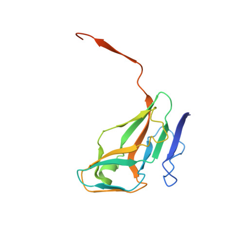
    dUTPases are a ubiquitous family of enzymes that are essential for all organisms and catalyse the breakdown of 2-deoxyuridine triphosphate (dUTP). In Bacillus subtilis there are two homotrimeric dUTPases: a genomic and a prophage form. Here, the structures of the genomic dUTPase and of its complex with the substrate analogue dUpNHpp and calcium are described, both at 1.85 A resolution. The overall fold resembles that of previously solved trimeric dUTPases. The C-terminus, which contains one of the conserved sequence motifs, is disordered in both structures. The crystal of the complex contains six independent protomers which accommodate six dUpNHpp molecules, with three triphosphates in the trans conformation and the other three in the active gauche conformation. The structure of the complex confirms the role of several key residues that are involved in ligand binding and the position of the catalytic water. Asp82, which has previously been proposed to act as a general base, points away from the active site. In the complex Ser64 reorients in order to hydrogen bond the phosphate chain of the substrate. A novel feature has been identified: the position in the sequence of the ;Phe-lid', which packs against the uracil moiety, is adjacent to motif III, whereas in all other dUTPase structures the lid is in a conserved position in motif V of the flexible C-terminal arm. This requires a reconsideration of some aspects of the accepted mechanism.


  • Organizational Affiliation
    • Department of Chemistry, University of York, England.

Macromolecule Content 

  • Total Structure Weight: 102.27 kDa 
  • Atom Count: 7,407 
  • Modeled Residue Count: 774 
  • Deposited Residue Count: 864 
  • Unique protein chains: 1

Macromolecules

Find similar proteins by:|  3D Structure
Entity ID: 1
MoleculeChains  Sequence LengthOrganismDetailsImage
PROBABLE DEOXYURIDINE 5'-TRIPHOSPHATE NUCLEOTIDOHYDROLASE YNCF
A, B, C, D, E
A, B, C, D, E, F
144Bacillus subtilisMutation(s): 0 
EC: 3.6.1.23
UniProt
Find proteins for O31801 (Bacillus subtilis (strain 168))
Explore O31801 
Go to UniProtKB:  O31801
Entity Groups
Sequence Clusters30% Identity50% Identity70% Identity90% Identity95% Identity100% Identity
UniProt GroupO31801
Sequence Annotations
Expand
Reference Sequence

Small Molecules

Ligands 3 Unique
IDChains Name / Formula / InChI Key2D Diagram3D Interactions
DUP

Query on DUP


Download:Ideal Coordinates CCD File
G [auth A]
L [auth B]
N [auth C]
P [auth D]
U [auth E]
G [auth A],
L [auth B],
N [auth C],
P [auth D],
U [auth E],
Y [auth F]
2'-DEOXYURIDINE 5'-ALPHA,BETA-IMIDO-TRIPHOSPHATE
C9 H16 N3 O13 P3
XZLLMTSKYYYJLH-SHYZEUOFSA-N
GOL

Query on GOL


Download:Ideal Coordinates CCD File
I [auth A]
K [auth A]
Q [auth D]
S [auth D]
V [auth E]
I [auth A],
K [auth A],
Q [auth D],
S [auth D],
V [auth E],
Z [auth F]
GLYCEROL
C3 H8 O3
PEDCQBHIVMGVHV-UHFFFAOYSA-N
CA

Query on CA


Download:Ideal Coordinates CCD File
H [auth A]
J [auth A]
M [auth B]
O [auth C]
R [auth D]
H [auth A],
J [auth A],
M [auth B],
O [auth C],
R [auth D],
T [auth D],
W [auth E],
X [auth E]
CALCIUM ION
Ca
BHPQYMZQTOCNFJ-UHFFFAOYSA-N

Experimental Data & Validation

Experimental Data

  • Method: X-RAY DIFFRACTION
  • Resolution: 1.85 Å
  • R-Value Free:  0.187 (Depositor), 0.201 (DCC) 
  • R-Value Work:  0.151 (Depositor), 0.170 (DCC) 
  • R-Value Observed: 0.153 (Depositor) 
Space Group: P 21 21 21
Unit Cell:
Length ( Å )Angle ( ˚ )
a = 99.116α = 90
b = 99.345β = 90
c = 99.252γ = 90
Software Package:
Software NamePurpose
REFMACrefinement
MOSFLMdata reduction
SCALAdata scaling
PHASERphasing

Structure Validation

View Full Validation Report


Ligand Structure Quality Assessment 


Entry History 

Deposition Data

Revision History  (Full details and data files)

  • Version 1.0: 2010-08-11
    Type: Initial release
  • Version 1.1: 2012-04-11
    Changes: Database references, Non-polymer description, Version format compliance
  • Version 1.2: 2023-12-20
    Changes: Data collection, Database references, Derived calculations, Other, Refinement description